Article Text

Statistics from Altmetric.com
Description
We present the case of a 74-year-old patient who, as an adult, enjoyed eating uncooked pork and beef. Recently, he had suffered from a sudden onset of gait disturbance, memory loss and disturbance of consciousness. He was brought to the emergency department for evaluation. On arrival, his vital signs were stable. The physical examination revealed mild weakness of the right extremities (muscle strength grade 3/5), slurred speech, left facial palsy and general appearance of weakness. ELISA was positive, as were serum and cerebrospinal fluid (CSF) parasite antibody immunoglobulin G for cysticercosis. We strongly suspected neurocysticercosis.
The brain CT scan (figure 1), brain MRI (figure 2), abdominal CT scan (figure 3) and plain X-rays (figures 4⇓⇓⇓⇓–9) had a characteristic ‘starry sky’ appearance, revealing calcified foci in muscles. Treatment of neurocysticercosis lesion includes administration of albendazole and steroids, and surgical ventriculoperitoneal shunting to alleviate the symptoms.

Brain CT revealing innumerable hyperdense ‘dot’ in the brain parenchyma.

Brain MRI showing that the numerous hypointense cysts are in the brain parenchyma.

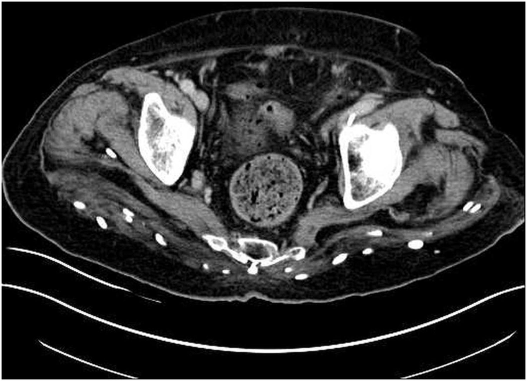
Abdominal CT revealing innumerable hyperdense ‘dot’ in the para lumbar spine and gluteal muscles.

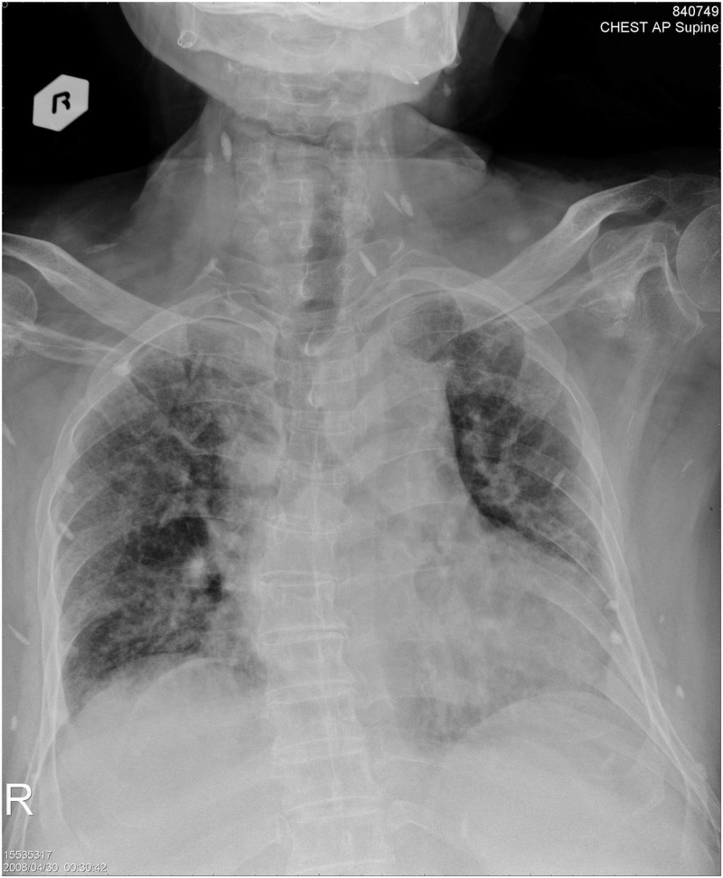
Chest film revealing numerous cysticerci on the neck and chest wall regions.
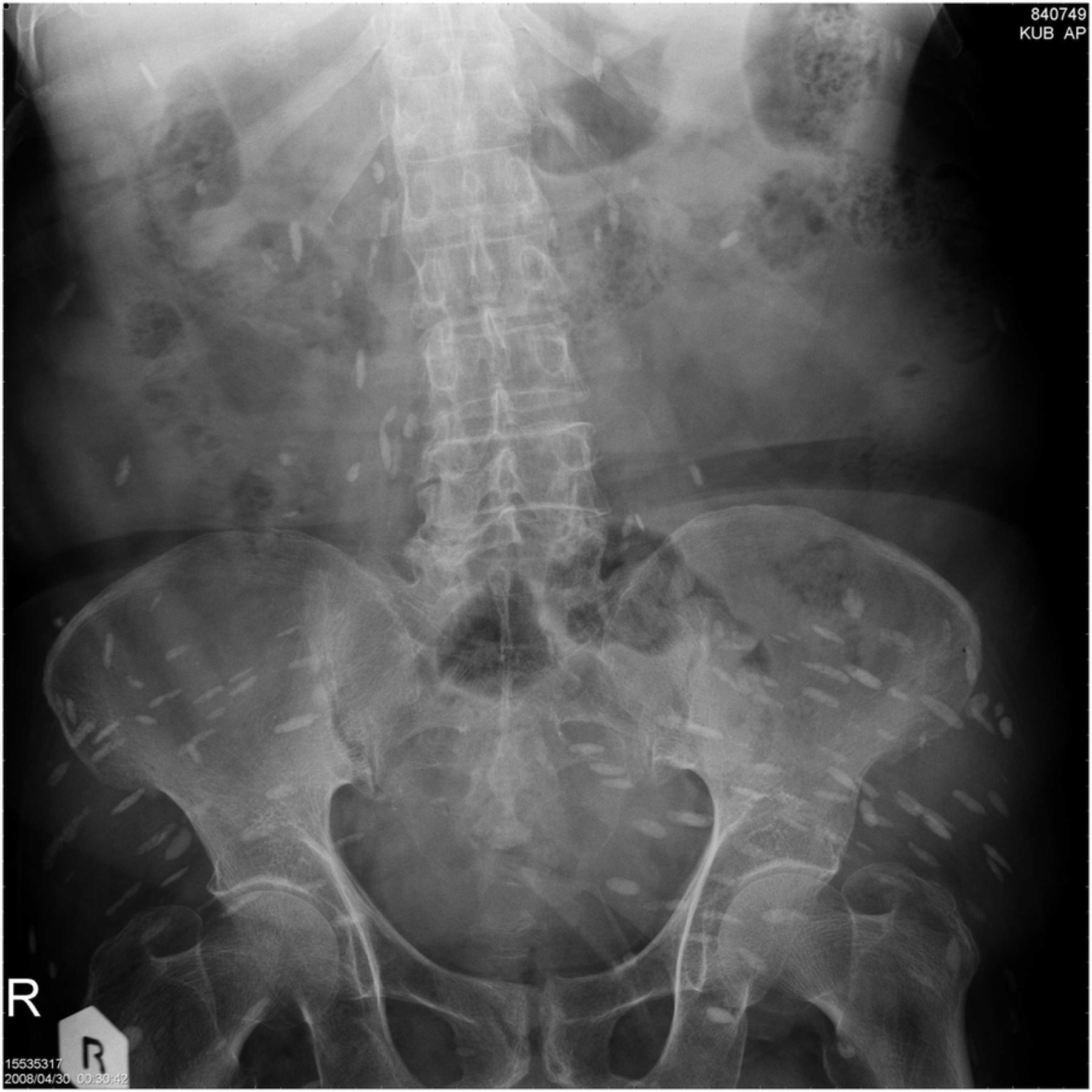
Kidneys, ureters, and bladder X-ray film revealing numerous cysticerci on the gluteal and iliopsoas muscle regions.
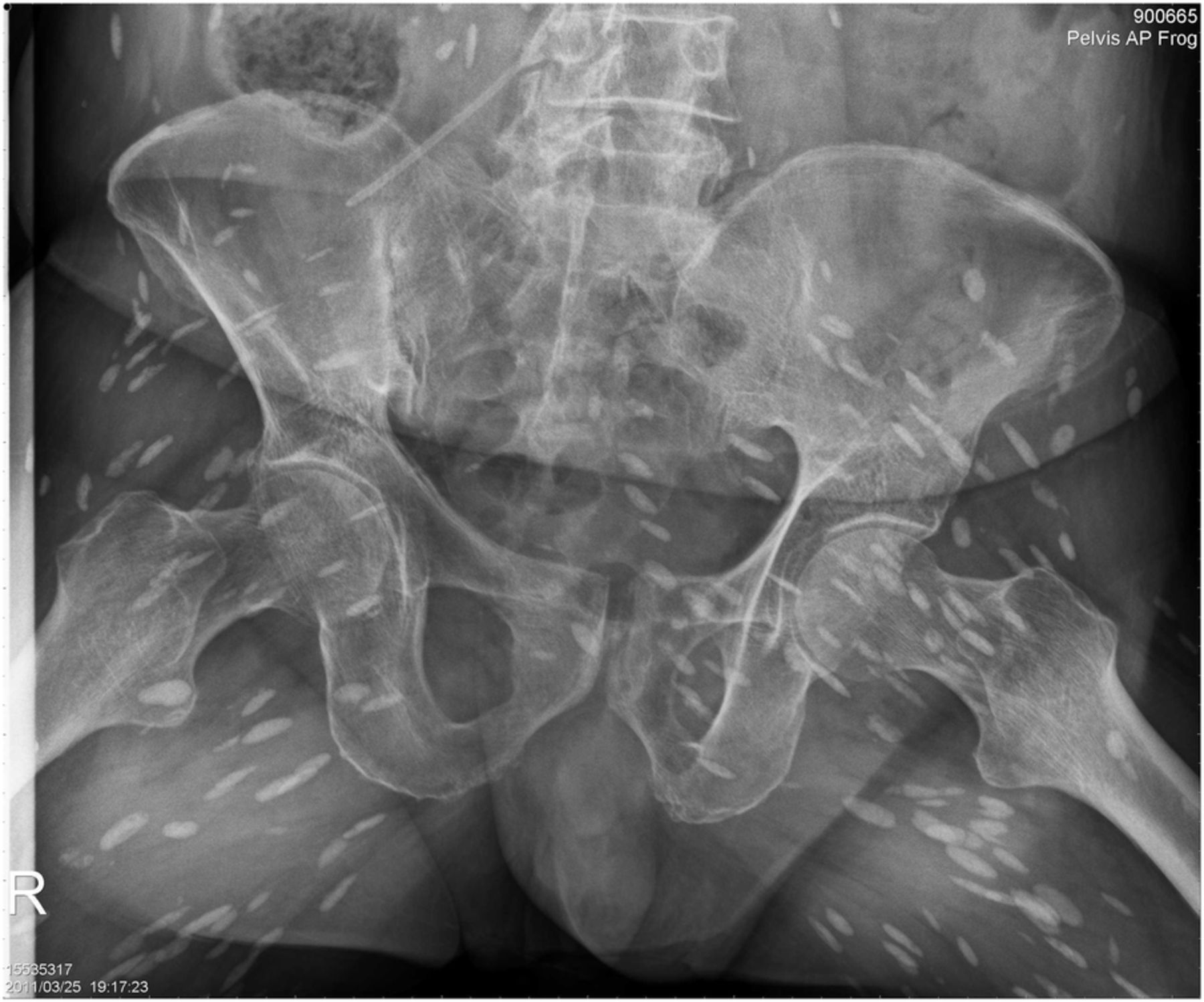
Pelvis film revealing numerous cysticerci on the gluteal and iliopsoas muscle regions.

Femur film revealing numerous cysticerci on the right thigh.

Knee film revealing numerous cysticerci on the left knee.

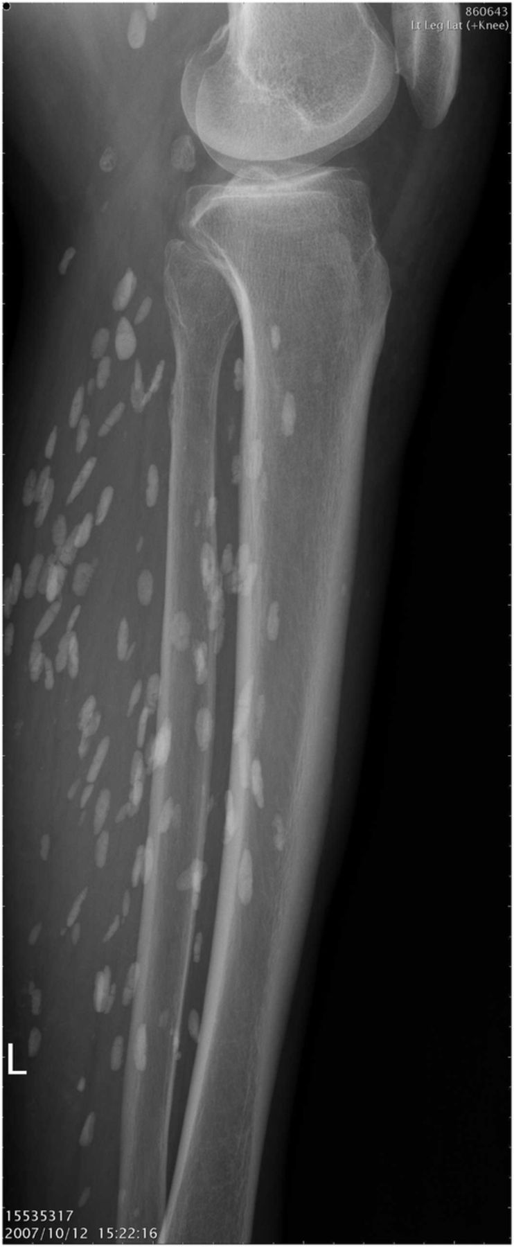
Leg film revealing numerous cysticerci on the left leg.
Disseminated cysticercosis is a very rare infectious disease.1 It is important to recognise disseminated cysticercosis clinically and to perform appropriate radiological investigations, because this condition requires an appropriate therapy. Patients who have not undergone treatment and who have active cysts remain at a risk of serious complications.
Learning points
-
Disseminated cysticercosis is a rare form of cysticercosis in which the cysticerci spread throughout the body.
-
It is important to recognise disseminated cysticercosis clinically and to perform appropriate radiological investigations, because this condition requires an appropriate therapy.
Footnotes
-
Contributors M-PL wrote the article, W-ST collated data and Y-LC revised the manuscript.
-
Competing interests None.
-
Patient consent Obtained.
-
Provenance and peer review Not commissioned; externally peer reviewed.