Article Text

Statistics from Altmetric.com
Description
A 38-year-old woman presented with long-lasting dull pain that she localised in her left hip. X-ray and MRI of the hip showed a small calcification of the labrum (a ring of cartilage surrounding the acetabulum) and excluded the suspected femoroacetabular impingement (where there is abnormal contact between the acetabulum and the femoral head-neck junction).1 However, X-ray incidentally detected two teeth in the left pelvis (figure 1A, anteroposterior view; B, oblique). In the presented MRI (figure 2) the caudal tooth is outlined and an arrow indicates its central pulp. Both teeth belong to a well-demarcated round tumour of 4.5 cm diameter that contains much fat. This fat appears dark due to the fat-suppression of the applied proton density-weighted turbo spin-echo sequence. The tumour originates from the left ovary (figure 2, arrowheads), consistent with an ovarian dermoid cyst. After some time of consideration the woman finally decided for laparoscopic ovarian cystectomy. Histopathology confirmed an ovarian dermoid cyst and detected no malignant tumour components.
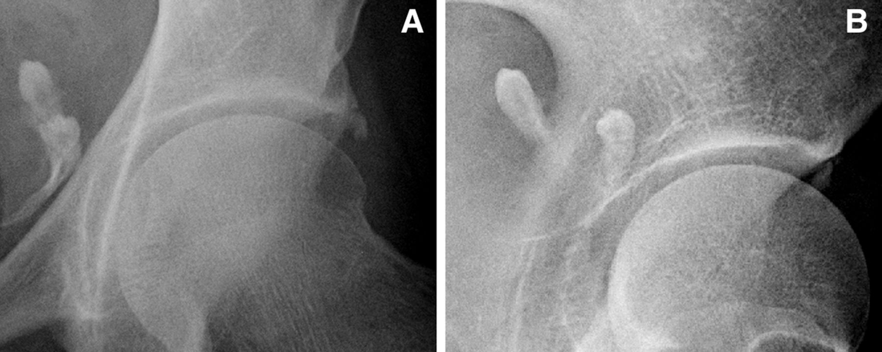
X-ray of the left hip (A, anterioposterior; and B, oblique) incidentally showed two teeth in the pelvis.

MRI showed a round tumour (*) originating from the left ovary (arrowheads). One of both teeth is outlined with its central pulp indicated by an arrow.
Ovarian dermoid cysts are the most common ovarian neoplasms. They are mature teratomas arising from the germ cells, and can therefore contain elements of all three germ cell layers such as epidermis, hair, calcified bone, teeth, fat and soft tissue.2 ,3 Often the tumour is asymptomatic and is detected incidentally, or is associated with unspecific symptoms. Over lifetime, malignant transformation occurs in about 1–2% of cases, mostly towards squamous cell carcinoma.3 ,4 This can be prevented by ovarian cystectomy.
Learning points
-
An ovarian dermoid cyst is a mature teratoma arising from the germ cells.
-
Often it is asymptomatic and is incidentally detected, for example, by teeth on a pelvic X-ray.
-
Malignant transformation (about 1–2% of cases) can be prevented by ovarian cystectomy.
Footnotes
-
Contributors JM and AS contributed to conception and design of the work, interpreted clinical data, drafted the article and revised it critically for important intellectual content, and approved the final version to be published. JM is the guarantor of the study and responsible for the overall content.
-
Competing interests None.
-
Patient consent Obtained.
-
Provenance and peer review Not commissioned; externally peer reviewed.