Article Text

Statistics from Altmetric.com
Description
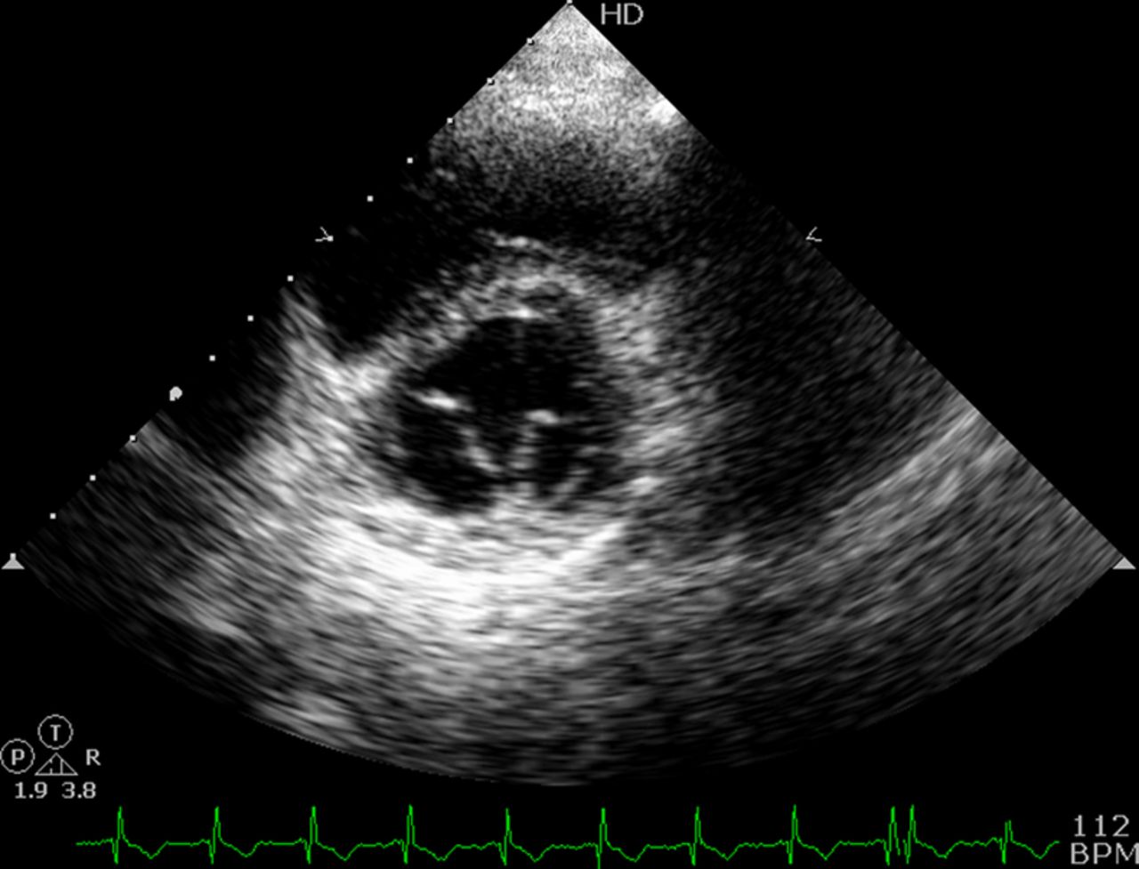
A 23-year-old asymptomatic man was referred for cardiac evaluation. A clinical examination was normal. Transthoracic 2D echocardiography showed double-orifice mitral valve (DOMV) in parasternal short-axis view at the level of mitral valve (figure 1, video 1). Apical four-chamber view showed a normally functioning valve without stenosis and/or regurgitation (video 2). Suprasternal long-axis view showed no evidence of coarctation (video 3). Rest of the echocardiographic examination was normal.

Parasternal short-axis view at the level of mitral valve showing two separate orifices of the double-orifice mitral valve.
Parasternal short-axis view at the level of mitral valve showing two separate orifices of the double-orifice mitral valve.
Apical four-chamber view with colour Doppler at the mitral valve showing competent and non-stenotic mitral valve.
Suprasternal long-axis view showing absence of coarctation.
Greenfield reported the first case of DOMV in 1876.1 It is characterised by the presence of two separate orifices in the mitral valve, each having independent chordal attachment to the papillary muscles at times creating an appearance of double parachute mitral valve. It is an extremely rare congenital anomaly which is usually found in association with other congenital heart diseases especially atrioventricular septal defect (most common), coarctation of aorta, interrupted aortic arch, patent ductus arteriosus and ventricular septal defect. Isolated DOMV, without other associated defects, is an extremely rare anomaly and the exact incidence is not known. It can present as mitral regurgitation (most common), incidental finding or uncommonly mitral stenosis.2 Abnormalities of the subvalvular apparatus are almost always present. It has been classified by Trowitzsch et al3 in 1985 into three types: complete bridge, incomplete bridge and hole type. DOMV usually comes to attention as an incidental finding with other congenital defects. However, isolated DOMV can be easily missed in apical four-chamber and parasternal long-axis views. Parasternal short-axis view is the best to diagnose this entity and should be carefully interrogated even in the absence of other defects.
Learning points
-
Isolated double-orifice mitral valve without other associated defects is an extremely rare anomaly which may be diagnosed as an incidental finding.
-
Comprehensive echocardiographic examination should be performed as it can be easily missed in most of the echocardiographic views.
-
Parasternal short-axis view is the best view to diagnose the anomaly.
Footnotes
-
Competing interests None.
-
Patient consent Obtained.
-
Provenance and peer review Not commissioned; externally peer reviewed.