Article Text

Statistics from Altmetric.com
Description
A gentleman treated 6 years ago for colonic carcinoma (stage-IIA) had been on regular follow-up for a span of 4 years, when he was apparently disease-free. After being lost to follow-up in the past 2 years, he was recently diagnosed with a pathological fracture of the seventh thoracic vertebra (figure 1) for which he underwent surgery (vertebral fixation and cord decompression) on an emergency basis.
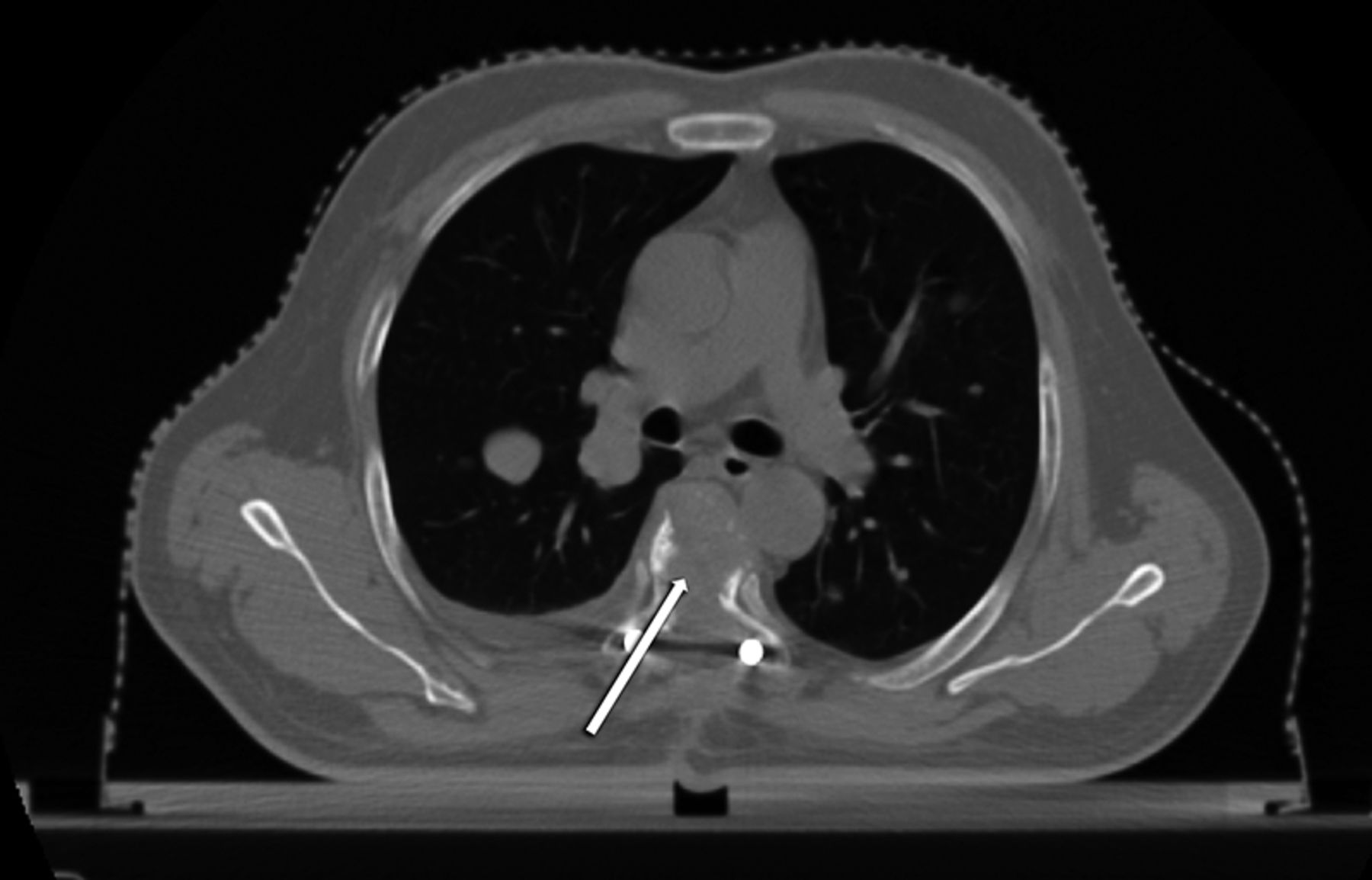
CT scan demonstrating the fractured seventh thoracic vertebral body (white arrow).
Postoperatively, he underwent a CT simulation of the thorax with the purpose of planning radiotherapy (RT) to the involved vertebrae. The CT-scan demonstrated multiple bilateral pulmonary nodules (figure 2). After palliative RT to the involved vertebral column, in the interest of academics, we utilised the CT-scan data for volumetric reconstructions to attempt a better depiction of the well-known phenomenon of ‘cannon ball metastases’.1 ,2 This was achieved by the use of Slicer 4.2, an open source software package (distributed under a BSD-style open source license) intended for advanced visualisation and analysis of medical image datasets.3 This, to our knowledge, is possibly the first publication providing a three-dimensional depiction of ‘cannon ball’ pulmonary metastases (figure 3).

Multiple, bilateral pulmonary nodules (‘cannon ball’ metastases) on traditional axial and coronal slices.

Three-dimensional rendition generated out of the thoracic CT-scan data, demonstrating ‘cannon balls’ within the lungs.
Slicer 4.2 is free to download and is available for use with the Linux, Mac and the Windows operating systems.4 While the package already contains numerous features, additional functionality can be via the in-built ‘extension manager’, which provides options to integrate various extra features. Though it is not currently approved for clinical use, it can, however, be used freely for academic research.
Learning points
-
The importance of adherence to follow-up remains undiminished even after initial years of uneventful follow-up, regardless of the stage of cancer.
-
Volumetric imaging data from sectional imaging can be digitally reconstructed to provide enhanced visualisation of normal and pathological anatomy.
-
Slicer 4.2 can be downloaded freely from http://www.slicer.org/pages/License, and being a feature-rich open source alternative to expensive commercial software, it can handle complex functions such as tractography, multivolume processing, etc.
-
Though not approved for clinical use, Slicer 4.2 can freely be utilised for research purposes.
Footnotes
-
Competing interests None.
-
Patient consent Obtained.
-
Provenance and peer review Not commissioned; externally peer reviewed.