Article Text

Statistics from Altmetric.com
Description
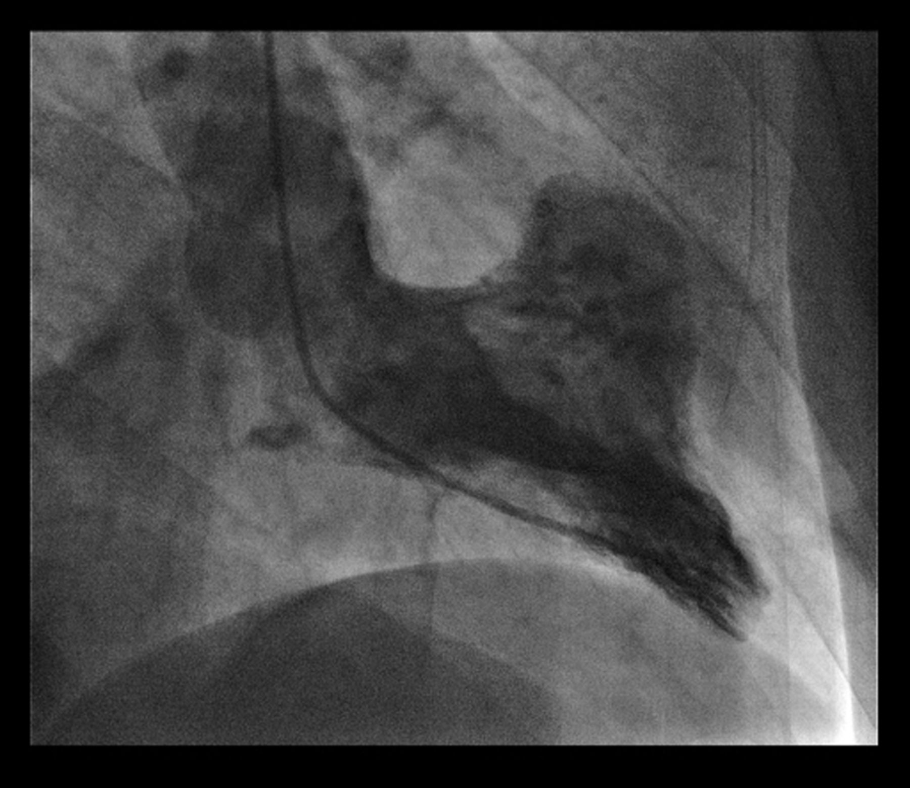
A 54-year-old man with typical ischaemic chest pain underwent emergency percutaneous coronary intervention for anterolateral ST elevation myocardial infarction. At this time, acute occlusion of an end artery, the intermediate branch of the left coronary artery, was identified (figure 1). A ventriculogram, right oblique view, also demonstrated dilation of the myocardium within the territory of the intermediate vessel (figure 2). At first, this was thought to be a cardiac diverticulum. The patient underwent coronary artery bypass grafting, and the diverticulum was, in fact, found to be a 3×3 cm thin-walled ventricular aneurysm. This was successfully repaired.

Coronary angiogram showing severe occlusion of the left main stem coronary artery and a small intermediate branch which is acutely occluded.
Right lateral oblique ventriculogram demonstrating left-ventricular aneurysm at the base of the heart in the territory of the intermediate vessel.
Left-ventricular aneurysms are a well-documented complication of myocardial infarction. Myocardial infarction accounts for 95% of cases.1 Usually, they are anterior-apical or inferior in origin, but rarely can occur elsewhere,1–3 and often co-exist with coronary artery diease.3 We report a rare basal left-ventricular aneurysm due to occlusion of an end artery, an intermediate branch of the left coronary arterial supply.
At the time of surgery, the defect wall was found to be extremely thin, conveying a risk of fatal left-ventricular free-wall rupture. Basal ventricular aneurysms are rare and provide a diagnostic dilemma,1 as seen in this case. Although not all aneuryms require surgical correction,1 basal aneurysms convey a much greater risk of rupture owing to the thinner myocardial wall in this region, and we therefore advocate repair in these cases. The patient had an uneventful postoperative recovery. Follow-up echocardiography at 3 months demonstrated no residual aneurysm.
Learning points
▶ Left-ventricular aneurysm (LVA) is a well-recognised entity occurring after myocardial infarction. The condition carries a significant morbidity and mortality and, under acute circumstances, may need surgical correction.
▶ Not all LVAs require surgical intervention, but the underlying coronary artery disease is often present which may require coronary artery bypass grafting.
▶ Basal aneurysms are extremely rare and they pose a diagnostic dilemma.
▶ Lack of adequate coronary collateralisation, together with left main stem disease, may produce ‘end artery’ necrosis, whereby acute ostial occlusion leads to localised myocardial necrosis and subsequently localised aneurysms.
▶ Basal aneurysm formation may be a direct indication of surgical repair because the myocardial wall in the area is extremely thin and the risk of rapture is high.
Footnotes
-
Competing interests None.
-
Patient consent Obtained.
-
Provenance and peer review Not commissioned; externally peer reviewed.