Article Text

Statistics from Altmetric.com
Description
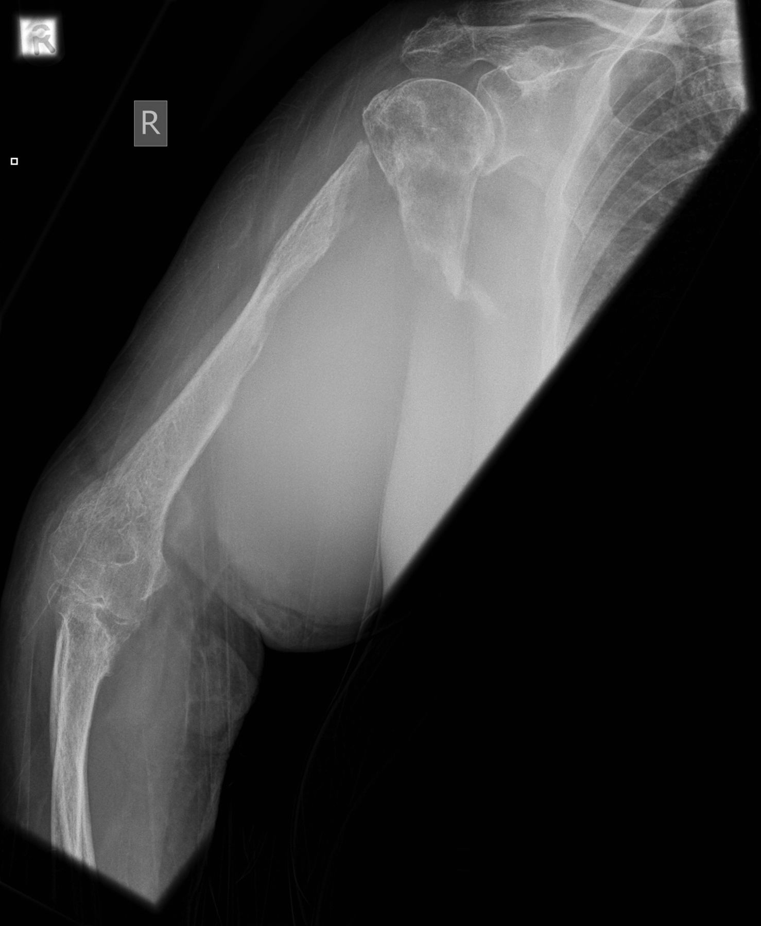
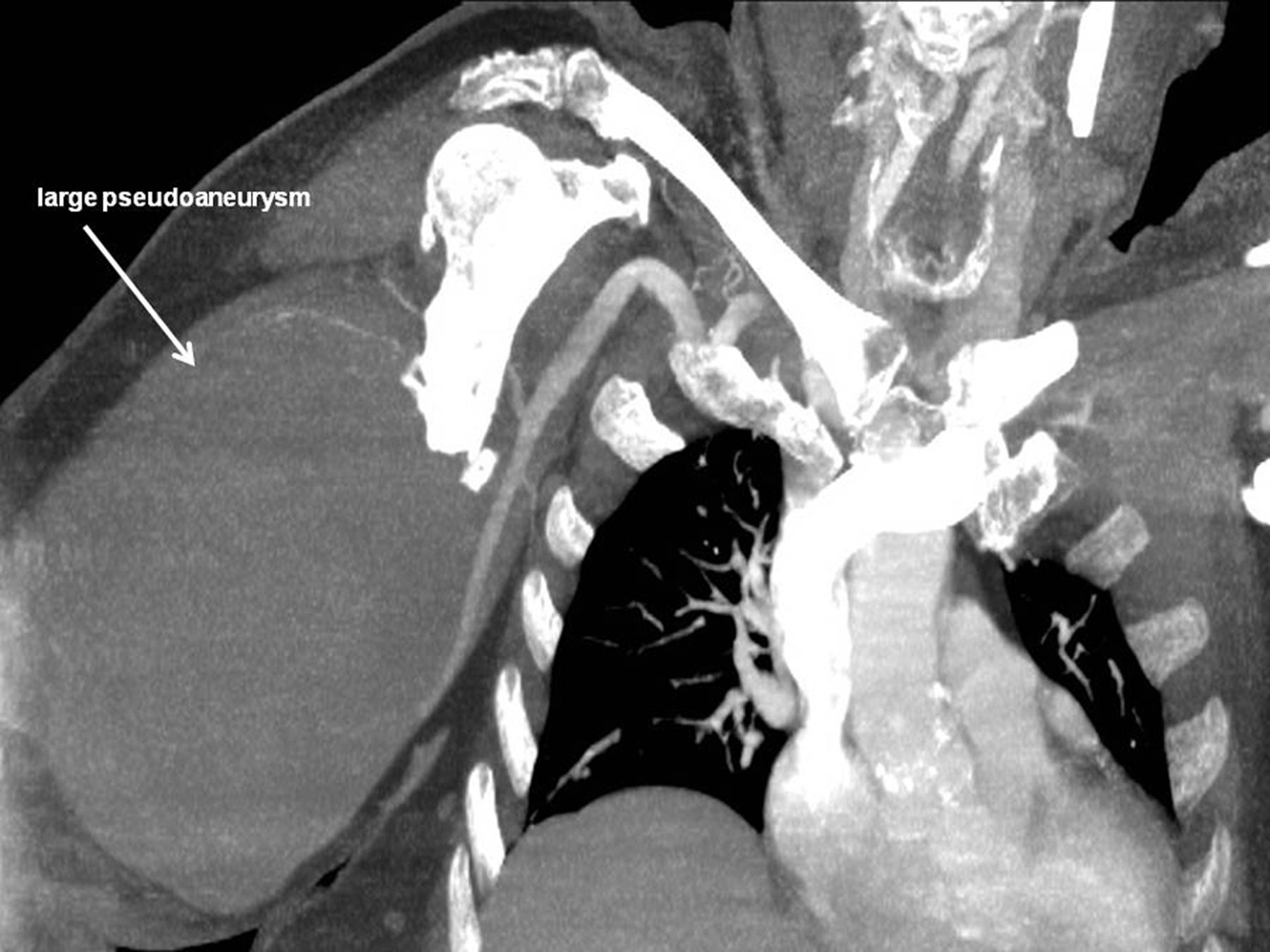
An 81-year-old woman presented with a right mid-shaft closed humerus fracture (dominant limb) following a mechanical fall. Her medical history included type 2 diabetes, hypertension, asthma and hypothyroidism. The patient was fully independent in all activities of daily living. Humeral fracture was managed conservatively, and the patient was discharged with a routine fracture clinic follow-up. Unfortunately, two consecutive follow-up appointments were missed by the patient and the first review after a fall was done with a delay of 9 months. During an outpatient visit, the patient complained of a chronic swelling, pain and general lack of function in the upper limb. Routine plain x-ray showed a chronic non-union of the humerus (figure 1). Examination revealed a swollen right upper limb and a mid-arm pulsatile mass with a systolic bruit on auscultation (figure 2). There was no evidence of vascular deficit. However, there was an obvious wrist drop with the reduced sensation in the radial nerve distribution and severely reduced range of all movements (figure 2). Clinical diagnosis of a flail upper limb secondary to brachial artery (BRA) pseudoaneurysm was made. This was confirmed by CT angiography (CTA), which demonstrated a 14 cm pseudoaneurysm arising from the defect in the mid-portion of the BRA (figures 3 and 4). After the discussion at the vascular MDT meeting, it was decided that the endovascular approach was impossible owing to the size of the pseudoaneurysm and presence of the displaced non-united humeral fracture. Therefore, the patient underwent an above elbow amputation with the intraoperative axillary artery controlled via infraclavicular incision to avoid haemorrhage (figure 5). Overall, the patient made an uneventful postoperative recovery.

Anteroposterior x-ray of the right shoulder demonstrating chronic non-union of the proximal humerus.

The patient with severe arm swelling and wrist drop.

CT angiography (axial view), which shows large pseudoaneurysm.

CT angiography (coronal view) presenting a large pseudoaneurysm in the arm.

The patient after an above elbow amputation.
Brachial artery pseudoaneurysms can be caused by trauma or iatrogenic interventions including brachial artery puncture during cardiac catheterisation or arterial gas sampling.1–4 These aneurysms usually present weeks to months after trauma. Delayed treatment and ongoing enlargement of the pseudoaneurysm can lead to life-threatening haemorrhage, venous oedema or compression of the neurological structures.5 Therefore, routine follow-up and the early diagnosis are crucial in avoiding a major limb amputation, which has profound psychological and physical consequences.
Imaging has an important role in the assessing of size and anatomical topography of the pseudoaneurysm. Different imaging modalities can be used including Doppler ultrasound, digital subtraction angiography and CTA like in the presented case. The goal of the treatment is to preserve the limb, which is achieved by exclusion of the pseudoaneurysm from the circulation, with or without the arterial reconstruction.6–8
The treatment algorithm depends on the size, location and accessibility of the pseudoaneurysm.3 Small pseudoaneurysms can be treated with a non-invasive procedure such as ultrasound-guided compression (USGC) during which the pressure is applied with an ultrasound transducer over the neck of the pseudoaneurysm until blood flow is eliminated.1 ,3 ,6 Unfortunately, the procedure is painful and often requires repeating for a considerable amount of time.3 Furthermore, USGC is associated with the high failure and recurrence rates.1 ,9 Promising results were reported with the use of percutaneous thrombin injection with the success rate as high as 90%.1 ,3 ,9 However, in a view of the limited number of reported studies such findings should be interpreted with caution.
Larger pseudoaneurysms require an invasive treatment by an open surgery. If the arterial defect is small it can be primarily repaired with sutures or a patch angioplasty.1 ,3 When this straightforward approach is deemed unfeasible, there are few alternatives. One of the surgical options includes an axillo-radial or axillo-ulnar bypass graft with the reversed great saphenous vein.3 ,6–8 This can be performed with or without the ligation of pseudoaneurysm (when pseudoaneurysm is thrombosed).10 The other alternative involves an excision of the pseudoaneurysm sac. The vascular continuity is then maintained by an end-to-end anastomosis between the interposition vein bypass graft and BRA.7 ,9 ,11
Additionally, some authors recommend the use of endovascular polytetrafluoroethylene (PTFE)-covered stent grafts, which were successfully used in the treatment of various peripheral pseudoaneurysms.6 Although PTFE stent grafts may be applicable in selected cases, lack of long-term results makes it difficult to draw definitive conclusions.6 ,10 ,7 Embolisation is primarily reserved for pseudoaneurysms arising from the small BRA branches.6 On the contrary, if the collateral circulation provides sufficient blood supply to the distal tissues, the embolisation of the main vessel (BRA) can be performed.10 Otherwise, the procedure carries the same rate of the limb loss (more than 50%) as the ligation of the BRA. However, it is essential to remember that ligation of the BRA at the expense of the limb loss can be a live saving procedure in cases of a ruptured pseudoaneurysm.10 Therefore, the decision regarding the treatment choice, with or without revascularisation procedures, needs to be balanced against the risks and tailored accordingly to the patient's needs.
Learning points
-
Patients with fractured humerus should be instructed about the importance of follow-up.
-
Brachial pseudoaneurysm can be easily identified on clinical examination as pulsatile mass with a systolic bruit on auscultation.
-
Management of the brachial artery pseudoaneurysm depends on size, location and functional state of the upper limb.
Footnotes
-
Competing interests None.
-
Patient consent Obtained.
-
Provenance and peer review Not commissioned; externally peer reviewed.