Article Text

Statistics from Altmetric.com
Description
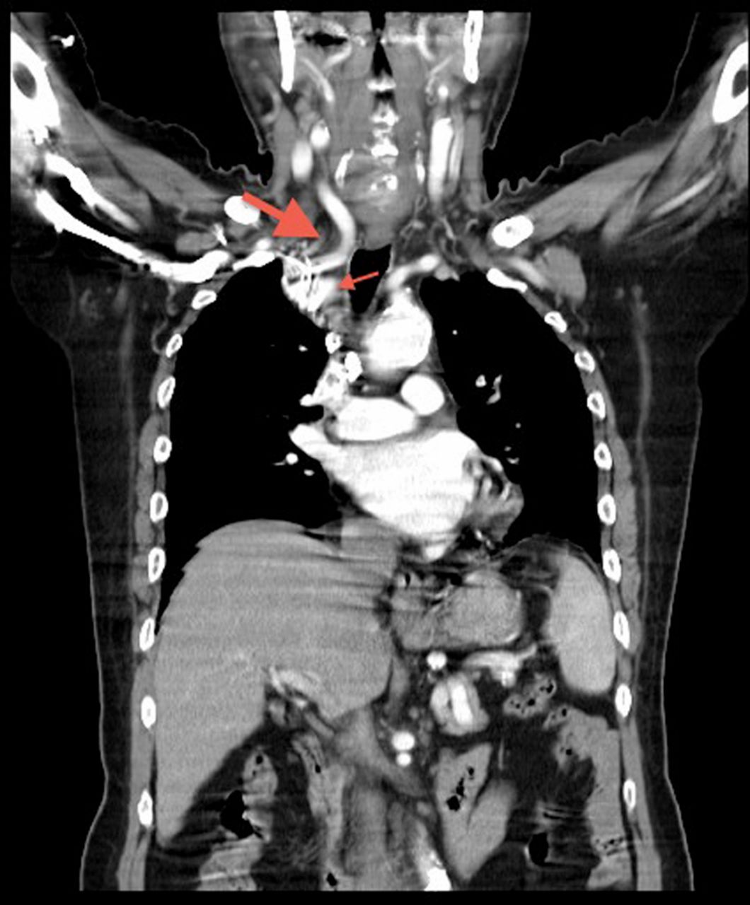
A 72-year-old man reported tarry stool for a day in the emergency department of a hospital. It was diagnosed that he had colon adenocarcinoma, and underwent left hemicolectomy 3 years ago. He did not receive any chemotherapy. A routine chest x-ray showed a round shadow in the upper lobe of his right lung (figure 1). He denied having fever, cough, shortness of breath or weight loss in the recent 2 months. Comparing the image with the one 2 years ago, we suspected that it might be recurrent colon cancer with metastasis of his right lung, enlarged mediastinal lymph nodes, lung tuberculosis, lymphoma or primary lung cancer. Surprisingly, the contrast-enhanced CT of chest revealed that this lesion was the shadow of distorted brachiocephalic artery and right common carotid artery (figure 2).

Chest x-ray: a round shadow in the upper lobe of right lung, near the clavicular head (red arrow).
Contrast-enhanced chest CT scan: the round shadow is composed of distorted right common carotid artery (big red arrow) and brachiocephalic artery (small red arrow).
When reviewing the chest x-ray again, we found that the latest one was anterioposterior view, and the patient rotated to the right side. This might result in magnifying the shadow of deformed vessels and make us misinterpret this image. In addition to chest CT scan, transthoracic sonography is another modality to investigate the suspected mass with wall contact in right upper lung.1 It is cheap, has no radiation exposure and easy access.2 The vessels can be distinguished easily from other solid tumour by colour Doppler sonography.
Learning points
-
Before you read a chest x-ray, be sure to determine whether it is qualified.
-
Sonograpy can be used to evaluate the suspected mass with wall contact in the lungs.
Footnotes
-
Competing interests None.
-
Patient consent Obtained.