Article Text

Statistics from Altmetric.com
Description
An 87-year-old woman presented to the emergency department with confusion, sinus tachycardia, hypotension and fever. Laboratory investigations confirmed metabolic acidosis, acute kidney injury (urea 18.5 mmol/l, serum creatinine 197 µmol/l and Egfr 21 ml/min/1.73 m2), depressed bone marrow function (platelets 136×109/l and white cell count (WCC) 3.0×10⁹/l) and elevated prothrombin and activated partial thromboplastin time (PT 15.8 s and APTT 61 s). Urinalysis was positive for blood, protein and leucocytes. Severe sepsis was diagnosed and treatment with intravenous fluids, co-amoxiclav and vitamin K was initiated. Proteus mirabilus was later identified from blood cultures.
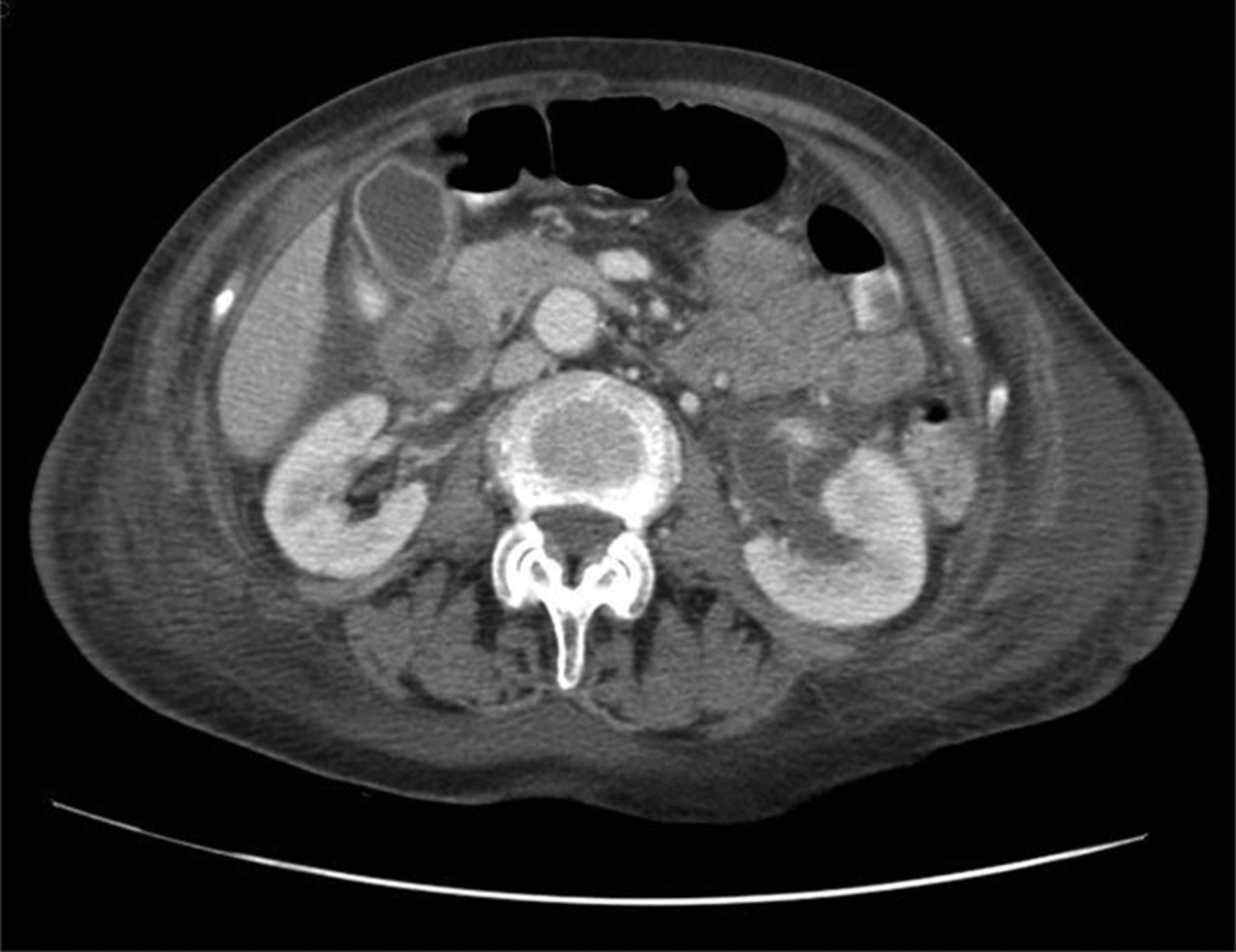
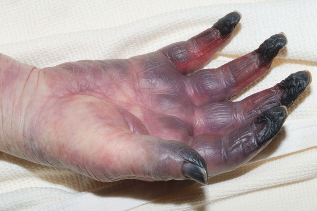
Despite treatment the patient deteriorated with abdominal tenderness, refractory hypoglycaemia, mucocutaneous bleeding, symmetric areas of petechiae, ecchymotic plaques and dry gangrene of the upper and lower extremities (figures 1 and 2). Laboratory investigations suggested disseminated intravascular coagulation (DIC) (platelets 13×109/l, PT 23.3 s, APTT 68.5 s and normal fibrinogen). A vasculitis screen was negative. She was treated with intravenous tazocin, hydrocortisone and platelets. On day 7, CT imaging demonstrated left pelvi-ureteric junction (PUJ) obstruction, rupture of the left renal tract (figure 3) and multiple abdominal and pelvic fluid collections. A left-sided ureteric stent was inserted by the urologists, and the digits affected by dry gangrene were autoamputated under the plastic surgeons.

Bluish discoloration and ecchymotic plaques of the palm and fingers with dry gangrene of the distal phalanges.
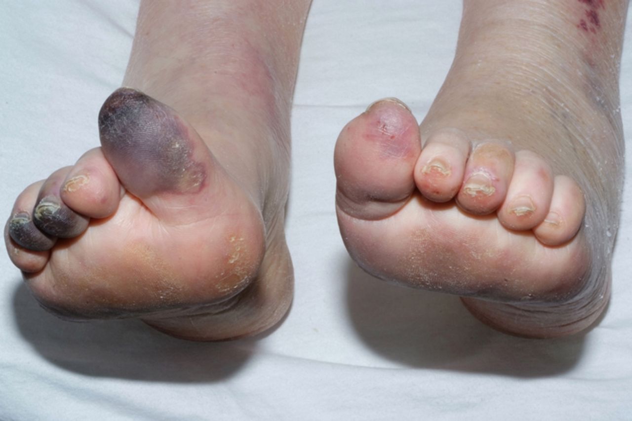
Petechiae, ecchymotic plaques and dry gangrene of the toes.
CT imaging of the abdomen demonstrating a fluid collection extending from the lower pole of the left kidney, continuous with a dilated proximal ureter, consistent with left PUJ obstruction.
Symmetrical peripheral gangrene (SPG) is a rare, but devastating complication of DIC, characterised by SPG with no evidence of large-vessel occlusion or vasculitis.1 Microorganisms commonly implicated include meningococci, pneumococci, streptococci and staphylococci.2 To our knowledge, this is the first reported case of SPB caused by proteus spp.3
Learning points
-
Symmetrical peripheral gangrene is a rare, but important, dermatological manifestation of disseminated intravascular coagulation.
-
Gram-positive microorganisms are most commonly implicated, but Gram-negative bacilli have been reported.
-
No treatment is universally effective and attention should be focused on correcting the underlying cause.
Footnotes
-
Competing interests None.
-
Patient consent Obtained.
-
Provenance and peer review Not commissioned; externally peer reviewed.