Article Text

Summary
Disorders of sex development (DSD) include congenital conditions where developments of chromosomal, gonadal or anatomical sex are atypical. Ostrer in 2000, reported a prevalence of 1:20 000 for 46 XY DSD and complete gonadal dysgenesis. A 21-year-old patient consulted for sexual ambiguity at the out-patient department of the Philippine general hospital. At birth, the perceived female external genitalia and clitoromegaly, led the parents to register and eventually rear the patient as a female. At puberty, he developed masculine features and growth of phallus. Patient was more interested in male activities and began to identify himself as male in the community. The discrepancy between his birth certificate and his male gender jeopardised his ambition to become a policeman; this led him to seek medical consult. On physical examination, he was phenotypically male. The external genitalia showed the phallus length of 3.5 cm and perineoscrotal hypospadias. Chromosomal sex was normal 46 XY with neither numerical nor structural aberrations in all cell lines, serum testosterone was low and gonadotrophins were elevated. Whole abdominal CT scan showed bilaterally undescended testes and a 4.5 cm blind vaginal pouch seen on genitogram. Bilateral orchidectomy with first stage repair of hypospadias was performed. On histopathology, the right testis was fibrotic and the left testis showed minimal testicular tissue with absent spermatids. The clinical, endocrine, cytogenetic and histopathologic data are consistent with gonadal dysgenesis syndrome.
Statistics from Altmetric.com
Background
The term disorder of sex development (DSD) includes congenital conditions in which development of chromosomal, gonadal or anatomical sex is atypical.1 DSD are a heterogeneous group of rare conditions. Thyen, Lanz, et al estimated a rate of 2.2/10 000 cases of ambiguous genitalia at birth with congenital adrenal hyperplasia as the most common cause of DSD in newborn.2 In Egypt Temtamy, et al reported an incidence of one newborn with ambiguous genitalia per 3000 live births.3 Hamerton, Canning, Smith, et al, reported an incidence of DSDs at 1:4500 to 1:5000live births.4 In this report, we present a case of 46 XY DSD. The clinical, endocrine, cytogenetic and histopathologic data are consistent with gonadal dysgenesis. Apart from the malignant potential of the dysgenetic gonads, the psychosocial impact of the disorder and its implications on late intervention must also be taken into account. This case report highlights the importance of early diagnosis for gender identity.
Case presentation
A 21-year-old phenotypical male presented at the general endocrinology clinic of the University of the Philippines–Philippine General Hospital in Manila, Philippines for the evaluation of the sexual ambiguity. He was the ninth in a family of fifteen, born by normal full-term delivery after an uneventful pregnancy. Apparently grossly female external genitalia with clitoromegaly were noticed at birth. No investigation for the clitoromegaly was done. Sex of rearing and psychological orientation was female. His mother had one abortion, but fourteen other siblings, of which are nine brothers and four sisters were normal. Parents were not related and of normal intelligence and stature. At 11 years of age, sexual hair development and phallic growth were observed. His physical and psychosocial orientation was geared towards male gender. He dressed up and identified himself as male in the community. He went to college and finished criminology, and would have wanted to become a policeman but failed in his medical exam when a discrepancy in his birth certificate and the gender he is identified with was noted. This scenario prompted the patient to seek medical evaluation. Physical examination revealed a normal upper/lower segment ratio. His height was 165 cm (within the 10th and 25th percentiles for normal Filipino male), and weight 54.0 kg. There was no axillary hair noted and no breast development. Pubic hair was abundant and exhibited an android distribution. Urological examination revealed ambiguous external genitalia. The phallus length was 4.0 cm covered by a redundant foreskin. The urethral opening was located in the perineum and there was no vaginal opening noted (figure 1). Labia majora were redundant, while labia minora were hypo plastic. There was no scrotum noted, but there was tenderness at the right inguinal area on minimal palpation. However, there were no palpable gonads at the inguinal canal and labia.

Physical examination findings: (A) upper left panel shows ambiguous genitalia. Note the phallus, male pattern pubic hair and absence of descended testes. Upper right panel shows a phenotypical male with no facial and axillary hair. (B) Lower left panel shows the perineal hypospadias and vaginal introitus. Lower right panel shows absence of breast development.
Investigations
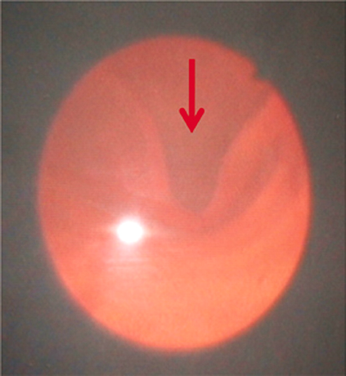
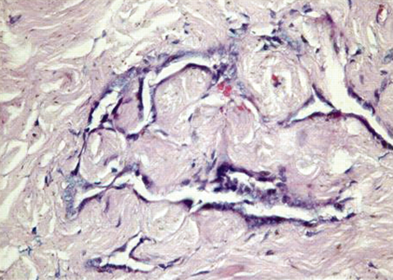
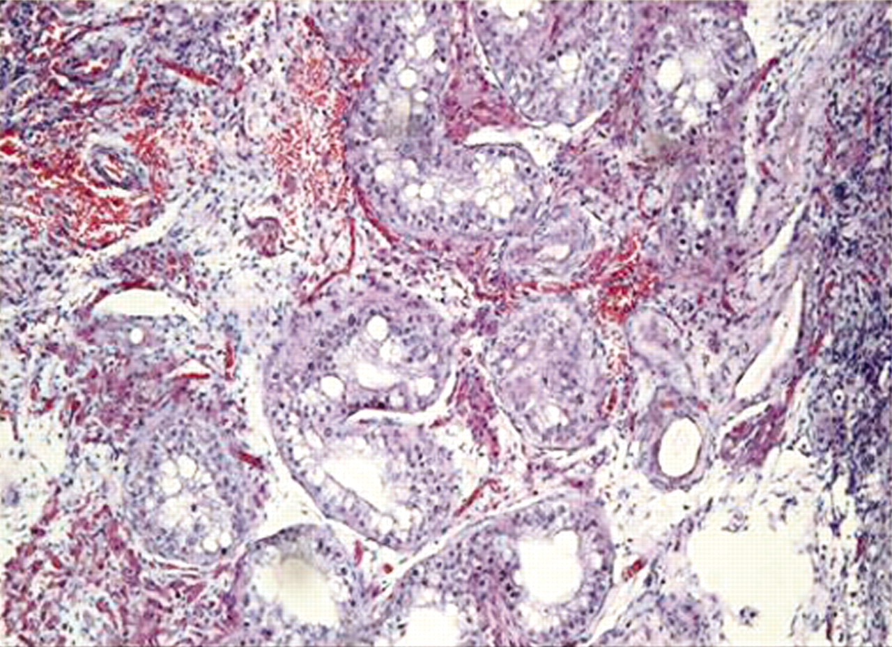
A CT scan of the whole abdomen disclosed the presence of undescended testis on the right, and another hypoechoic focus on the left pubic region, probably a testicular remnant. A genitoscopy was done and disclosed a blind ending vaginal pouch with a depth of 4.2 cm (figure 2). Cytogenetic studies revealed a normal 46, XY chromosome complement with neither numerical nor structural aberrations was found in all cell lines studied (figure 3). Hormonal assay showed low testosterone 5.4 nmol/l (9–38) with an elevated gonadotrophins follicle-stimulating hormone=34.4 mIU/l (1.0–10), luteinising hormone (LH)=13.8m IU/l (1.9–9.4). Other laboratory values were a normal 17-hydroxyprogesterone (17-OHP) at 1.1 nmol/l (normal <6) and a low dehydroepiandrosterone (DHEA) at 1.1 umol/l (normal 2.20–15.20) (table 1). After completion of the endocrinologic studies and imaging, a diagnostic laparoscopy was done and confirmed the bilaterally undescended testes. A genitogram was done and disclosed a 3.5 cm blind ending vaginal pouch (figure 4). Laparoscopic orchidectomy was carried out to remove the undescended testes. Major surgical findings noted grossly atrophied testes bilaterally. Repair of the hypospadias was also done in this patient. Histopathologic examination of the right testis (figure 5) showed that it was composed of fibrous tissue with collapsed cystic spaces representing testicular remnants, while the left testis (figure 6) showed paucity of testicular tissue with scarce Sertoli cells and no identifiable spermatids. These histopathology findings are consistent with testicular dysgenesis syndrome.
Summary of endocrine tests

Genitoscopy showing the opening of the blind-ending vaginal pouch.

The patient’s karyotype was that of a male showing a chromosomal complement of 46, XY.

Genitogram showing blind-ending vaginal pouch measuring 4.5 cm.

Microscopic examination of the right testis showed cystic spaces lined by low cuboidal to flattened epithelium, and surrounding fibrosis (H&E stain, 400x magnification).

Microscopic examination of the left testis showed testicular tissue composed mostly of tubules lined by Sertoli cells with no identifiable spermatids (H&E stain, 100x magnification).
Differential diagnosis
Other causes of undervirilisation in 46 XY DSD are represented by defects in androgen formation attributed to enzymatic defects along the conversion of cholesterol to testosterone, which occur due to autosomal recessive mutations. Patients with the more proximal enzymatic blocks in the cholesterol to testosterone pathway resemble congenital adrenal hyperplasia as well as 46 XY DSD in their presentation. These patients usually present early with severe salt wasting, and if not recognised and treated early, the condition may lead to death. However, these entities were ruled out in this patient due to normal 17 OHP, and abnormal histopath findings. There was also no history of adrenal insufficiency in our patient. On the other hand, the presence of atrophic/ rudimentary epididymes and the development of secondary male sexual characteristics and phallic growth at the onset of puberty indicated the production of testicular androgens. This endocrine and morphological feature rule out the presence of LH receptor defect as seen in the case of Leydig cell hypoplasia.
Treatment
The holistic management of DSD focuses on the medical – surgical and psychosocial issues.5 The gender identity if not addressed early in children and adults lead to psychosexual dilemma, emotional distress and even risk of social isolation, embarrassment and discrimination in the society. Our patient presented to the general endocrinology clinic for evaluation, only after being disqualified from the job he was applying for because of his physical condition. Thus, early recognition of this condition for proper gender assignment is important to avoid the psychosocial issues later in adulthood. One important universally applicable ethical principle is the consideration of a possible malignant potential of an undescended testis especially on a dysgenetic gonad. Because of this consideration, our patient underwent bilateral orchidectomy and first stage repair of the hypospadias. He is presently on hormonal replacement with testosterone injection and since patient was registered as female at birth–the legal issue of his gender change is also in the process.
Outcome and follow-up
In a review of literature, a patient with a Y chromosome has a 20–30% risk of developing a gonadoblastoma and/or invasive germ cell tumour in a streak or dysgenetic gonad.6 Apart from the malignant potential of the dysgenetic gonads, the psychosocial impact of the disorder and the implications of late intervention. The sensitivity of the subject means that children and adults with DSD are particularly vulnerable to psychosexual dilemma, emotional distress and even risk of social isolation, embarrassment and discrimination in the society. This case report further aims to highlight the importance of early diagnosis and decision–intervention on DSD patients.
Discussion
The case reported herein represents sporadic and incomplete form of the XY gonadal dysgenesis syndrome. This patient had normal female external genitalia at birth with clitoromegaly. During pubertal development secondary male sexual characteristics were noted and phallic growth was noticed. On investigation revealed no mullerian structures except for the blind ending vaginal pouch seen on genitoscopy being the remains of the urogenital sinus. This exemplifies that the tubular compartment of the dysgenetic testes was only partially damaged and the interstitial compartment was importantly impaired. The dysgenetic testes were capable of producing a sufficient amount of anti mullerian hormone for mullerian regression and testosterone for wolffian development, although they were incapable of producing sufficient amount of T for complete masculinisation of external genitalia. A detailed classification of XY gonadal dysgenesis was proposed by Kofman et al in his report elucidating comparison of the complete and incomplete forms of the syndrome.7 Overall data demonstrate that the phenotypic expression of the XY gonadal dysgenesis depends on the degree of damage in each of the testicular compartments.7
In two similar case reports on XY gonadal dysgenesis, (T) testosterone response to HCG stimulation was done to differentiate the various forms of gonadal dysgenesis. In this report, complete gonadal dysgenesis and type II (interstitial compartment) partial gonadal dysgenesis is associated with absence of T response to human chorionic gonadotropin (HCG) stimulation, whereas the type I (tubular compartment) is noted with rise in T on HCG stimulation.3 However, this was not done in our patient for practical reasons. The low basal T, low DHEA, elevated gonadotrophins, normal 17-OHP with histopathologic features characterised by paucity of testicular tissues, absence of spermatids, with only remaining fibrous tissue seen on the right testis is consistent with our diagnosis of XY gonadal dysgenesis.
Learning points
▶ Gonadal dysgenesis is one of the causes of ambiguous genitalia in a 46, XY patient.
▶ Gonads show characteristic abnormal development of the tubular and/or interstitial testicular compartments. It is hormonally characterised by low testosterone and elevated gonadotrophin levels.
▶ Sex assignment and gender of rearing during childhood in this case differed from the gender the patient preferred during adulthood, and created difficulties for the patient in applying for work.
▶ The management of DSD should not solely focus on medical–surgical issues but should also consider the condition’s social and emotional impact.
Footnotes
-
Competing interests None.
-
Patient consent Obtained.