Article Text

Statistics from Altmetric.com
Description
A 10-year-old female presented with history of prominent eyes since birth. She also gave history of deviation and occasional dislocation of left eye which she is able to retroplace on her own. Bilateral proptosis with large degree of exotropia left eye was seen in primary gaze. She had normal intellectual capacity with vision of 20/50 in right eye and Counting Fingers (CF) in left eye. She had midfacial hypoplasia, hypertelorism and dental abnormalities (Figure 1–3). Her cornea had no signs of exposure keratitis and fundus did not reveal any abnormality.
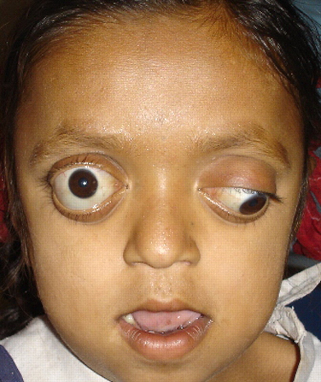
Anterior view of a 10-year-old female with Crouzon syndrome. Note the acrocephaly, proptosis, strabismus, hypertelorism, parrot-beaked nose and maxillary hypoplasia characteristic of this syndrome.

Oblique view: shallow orbit with midfacial hypoplasia.

Dental deformities.
Craniosynostoses are group of rare hereditary disorders characterised by premature closure of cranial sutures in the embryonic period or during early childhood. Crouzon syndrome is caused by premature closure of coronal and sagittal sutures. It has incidence of approximately 16.5 cases per million live births. The prevalence of this disease is 333–476 per million births.
Bilateral severe proptosis and corneal exposure are due to arrested growth of the maxilla and zygoma resulting in shallow orbits. Rarely the globe may be luxated anterior to the lids. Hypertelorism, strabismus (V pattern exotropia) and amblyopia are commonly found.
There may be associated chronic papilloedema causing optic atrophy. Other associations are aniridia, blue sclera, cataract, coloboma, ectopia lentis and optic nerve hypoplasia. No medical treatment exists for craniosynostosis. Surgical treatment in the form of cranial vault reconstruction should be done in the first few months of life.
According to Gray et al1 visual impairment in at least one eye occurred in 35% of patients and was bilateral in 9%. The most common cause of visual impairment was amblyopia hence amblyopia management should be a priority.
References
Footnotes
-
Competing interests None.
-
Patient consent Obtained.