Article Text

Statistics from Altmetric.com
Description
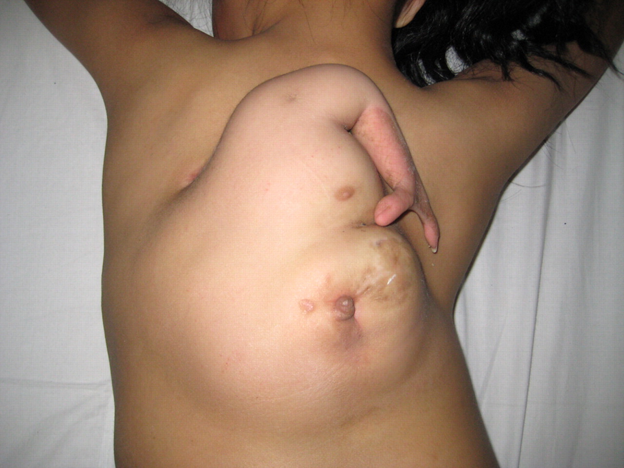
An 11-year-old girl was hospitalised because of a large mass in her back (figure 1). She was the second child in her family where there was neither twin pregnancy nor fetal malformation. The large mass has two parts where the upper part consists of a mammary gland and an attached upper limb including a scapular, humerus and two fingers, and where the lower part contains a thick-walled sac which endlessly secreted mucosal fluid. There was not a clear cleavage plane between the autosite and the parasite. The anteroposterior spine radiograph showed congenital scoliosis anomalies (figure 2), and the CT scan showed diastematorrhachia and diastematomyelia in the thoracolumbar region (figure 3). Based on above-mentioned physical examination and imaging findings, a diagnosis of fetus in fetu was thus made preoperatively. A surgical excision of the fetus in fetu was performed successfully. Histological sections of the excised sac specimen showed stratifyed squamous epithelium and digestive tract mucosa. Fetus in fetu is a very rare condition, with a reported incidence of one in 5000 000 live births.1 It is a malformed parasitic monozygotic diamniotic twin that is found inside the body of the living child or adult.2 Hoeffel et al2 provide an excellent and very complete literature review of 88 cases including their own a case. The mass is located in the retroperitoneum in 80% of cases. It also occurs in other locations, such as skull, sacrum, scrotum and mouth. However, it has never been reported in the back.

An 11-year-old girl had a large fetus in fetu in her back
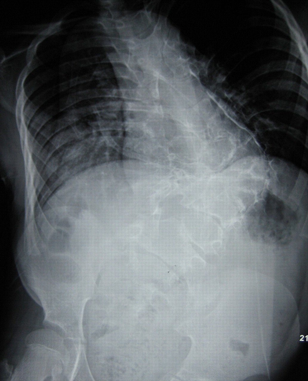
Anteroposterior spine radiograph showed congenital scoliosis anomalies

CT scan showed diastematorrhachia and diastematomyelia in the thoracolumbar region (arrow).
Learning points
Fetus in fetu is a very rare condition, with a reported incidence of one in 5000 000 live births.
It is a malformed parasitic monozygotic diamniotic twin that is found inside the body of the living child or adult. The mass is located in the retroperitoneum in 80% of cases. It also occurs in other locations, such as skull, sacrum, scrotum and mouth.
Fetus in fetu first was reported to occur in the back.
Footnotes
-
Competing interests None.
-
Patient consent Obtained.