Article Text

Statistics from Altmetric.com
Description
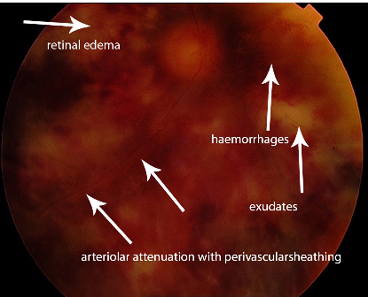
A 34-year-old lady who defaulted highly active antiretroviral therapy (HAART), returned after 2 years with bilateral rapidly progressive blurring of vision of 6 months duration. Five months back she was diagnosed to have cerebral toxoplasmosis and was started on double strength trimethoprim/sulphamethoxazole and HAART and she was continuing on the same regimen. Her CD4+T cell count was 147 (CD4+). Pupils were bilaterally dilated, sluggishly reacting to light. Visual acuity revealed perception of hand movements+ on the right eye and perception of light+ on the left eye. There was no focal neurological deficits. CT scan of brain showed a healed granuloma on the right side. With a past history of cerebral toxoplasmosis and CD4+ 147 (reconfirmed), the possibilities considered were toxoplasma choroidoretinitis or cytomegalovirus retinitis (CMV). But funduscopy revealed active retinitis bilaterally (figure 1A,B) with brush fire appearance, haemorrhages, exudates, arteriolar attenuation with peri vascular sheathing and retinal oedema diagnostic of CMV retinitis(figure 2). Deterioration of vision ceased once the treatment was initiated on intravenous ganciclovir.

(A, B) Fundal image showing active retinitis bilaterally.
Fundal image showing active retinitis brush fire appearance, haemorrhages, exudates, arteriolar attenuation with peri vascular sheathing and retinal oedema.
Learning points
Fundoscopic features are diagnostic of cytomegalovirus retinitis.1
CMV retinitis occurs with CD4+ less than 100 and in majority with CD4+ 50 or less but exceptions are documented in literature.2
In the HAART era, CMV retinitis may remain quiescent despite extremely low CD4 cell counts and rarely, CMV retinitis may become active in the setting of persistently high CD4 cell counts in a subset of HAART responders.2
There are reports in literature which mention that increased CD4 T lymphocyte counts may not protect against CMV retinitis.3
Footnotes
-
Competing interests None.
-
Patient consent Obtained.