Article Text

Statistics from Altmetric.com
Description
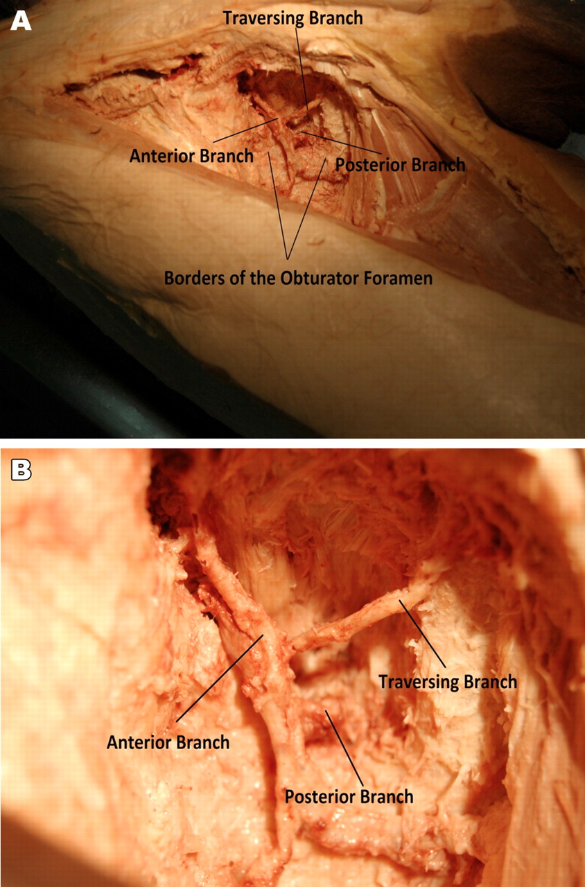
The variability of the obturator artery inside the pelvis is well known; however in the thigh only two branches encircling the obturator foramen are described. With the evolution of minimally invasive trans-obturator approaches to pelvic surgery the anatomy in this area needs to be better understood. Trans-obturator approach has been shown clinically more effective and less invasive treatment for urinary stress incontinence1 ,2; however it may be associated with vascular complications.3 To describe, confirm and measure the branches of the obturator artery following passage through the obturator foramen using a single human cadaver, dissection of the course of the obturator artery from its passage through the obturator foramen in the right groin was performed (figure 1). Vessels were measured with calipers and the branching patterns described. The obturator artery was identified piercing the obturator fascia at 7o’ clock position giving off posterior and anterior branches (2.7 mm and 1.9 mm in diameter, respectively) encircling the foramen measuring 2.7 by 5.2 cm. There was a traversing branch (1.7 mm in diameter) coming off the posterior branch 1.7 cm above the inferior border of the foramen. This branch passed the foramen medially 3.2 cm above the inferior border of the foramen. This study reveals that traversing branches of obturator artery are potentially at risk of injury during the trans-obturator surgical approach. Further dissections are required to understand how frequent this is.

Photograph of dissection illustrating the right femoral triangle (A) from a distance, (B) close-up.
Learning points
Traversing branches of obturator artery are potentially at risk of injury during the trans-obturator surgical approach.
Further dissections are required to understand how frequent this is.
Footnotes
-
Competing interests None.
-
Patient consent Not obtained.