Article Text

Statistics from Altmetric.com
Description
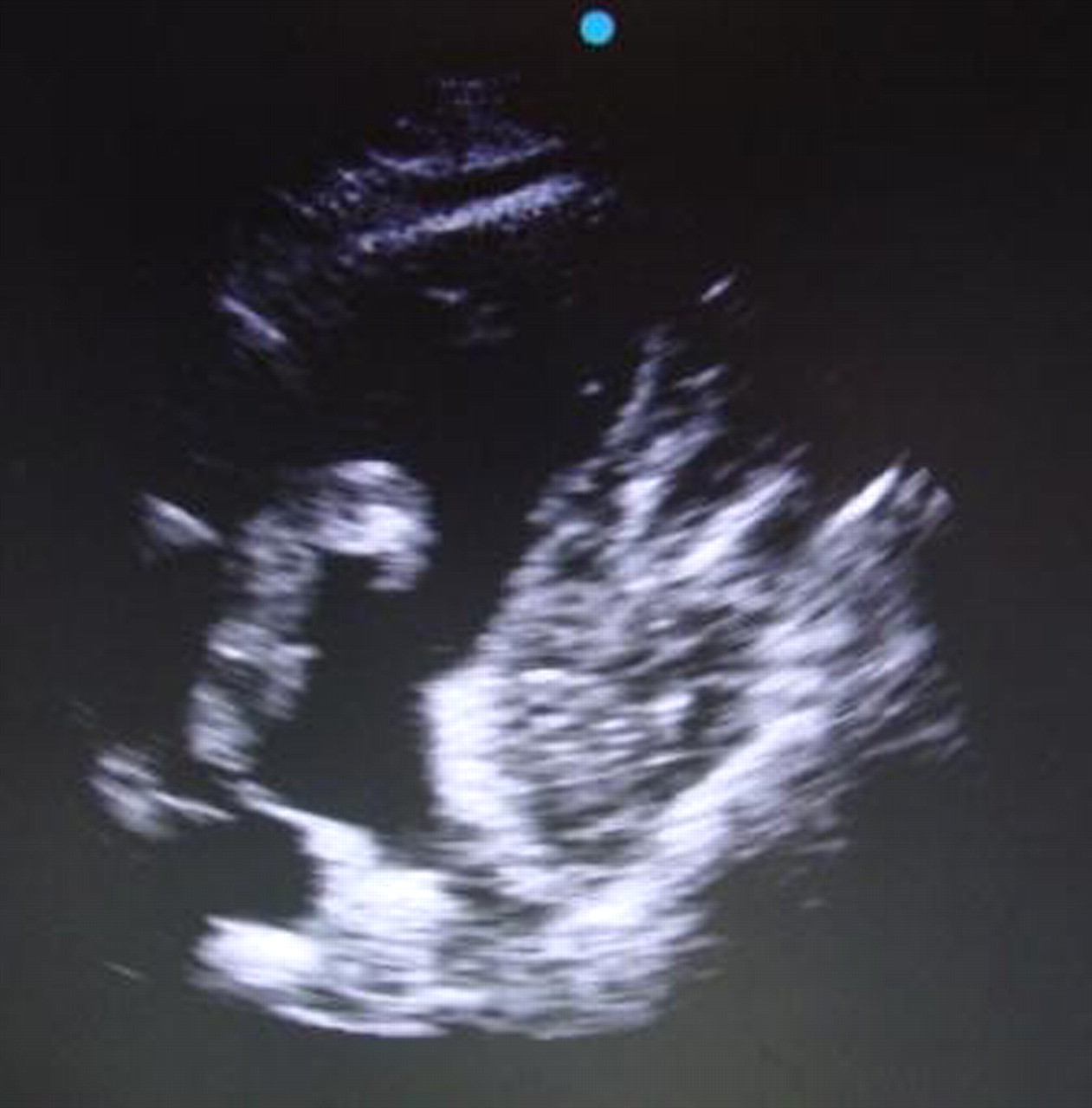
An 80-year-old man attended the emergency department (ED) after collapsing in church. When seen he was tachycardic, breathless and hypotensive. The ECG showed tachycardia. ED ultrasound showed an enlarged right ventricle with tricuspid regurgitation (video 1). Further examination revealed a lesion in the right atrium prolapsing into the right ventricle (figures 1 and 2 and video 2). Diagnosis of pulmonary embolism was made and the patient thrombolysed. Apart from age there were no obvious risk factors. Unfortunately despite treatment he deteriorated and later died. Peri-mortem imaging failed to show thrombus in the heart (figure 3), likely because it had passed into the pulmonary artery. Postmortem examination confirmed coiled, unorganised thrombus severely occluding both pulmonary arteries. ED ultrasound has been recommended in the evaluation of shock.1 Echocardiography is part of this and can exclude pulmonary embolism (PE) if right ventricular overload or dysfunction is absent. The presence of these features in shock with suspected PE may justify2 aggressive treatment. Elevated troponin 0.25 ng/ml (normal 0.00–0.1) with shock put this patient in the high risk category.2 Right heart thrombi are found almost exclusively in those with suspected or proven pulmonary embolism.1 Mortality, especially if free floating is high, irrespective of treatment and if untreated is associated with mortality of 80–100%.2 Immediate therapy is necessary, but optimal treatment controversial. Thrombolysis and embolectomy are probably both effective.1 3 This patient was highly unstable and further imaging or embolectomy unrealistic.
Tricuspid regurgitation.
Dilated right ventricle and echogenic thrombus in right atrium prolapsing into right ventricle.

Echogenic lesion seen within right atrium.

Thrombus seen prolapsing from right atrium into right ventricle.

Absence of echogenic lesion previously seen.
Learning points
ED echocardiography allows rapid diagnosis and treatment despite images not being ideal due to difficult positioning, lighting etc.
Footnotes
-
Competing interests None.
-
Patient consent Obtained.