Article Text

Statistics from Altmetric.com
Description
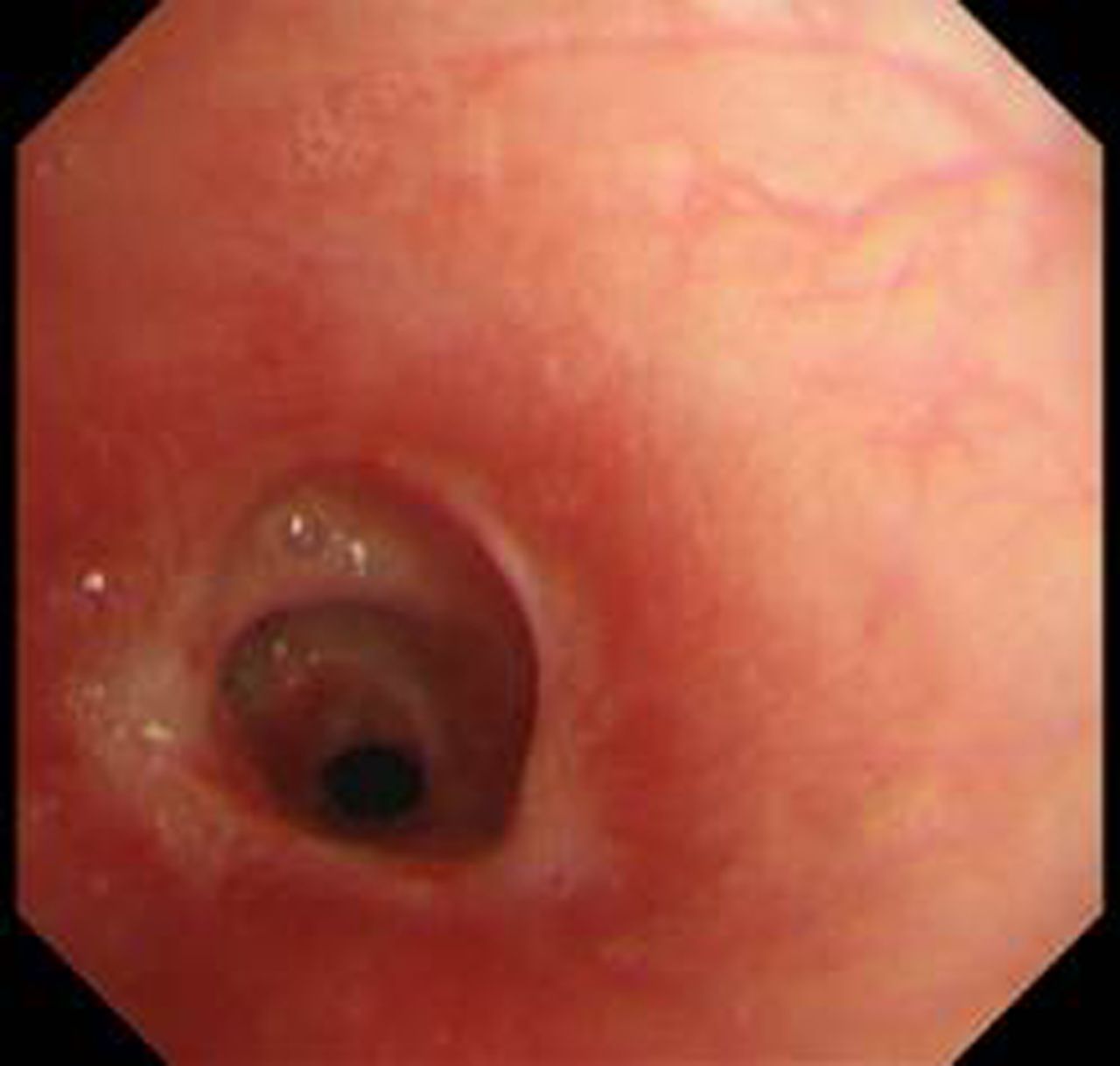
A 22-year-old woman was admitted to our hospital because of loss of consciousness. She was seen ingesting excess schizophrenia medication following an argument. Twelve hours later, she was found unconscious and surrounded by vomit. Assessment on admission revealed: Glasgow Coma Score E1V1M4, temperature 38.4°C and pulse rate 135 bpm. She was severely underweight, with a body mass index of 15. She was intubated without difficulty immediately after arrival using a 7 mm internal diameter endotracheal tube. Three days later, she was fully awake and was extubated uneventfully. Chest x-rays revealed infiltrative shadows in both lungs. She was diagnosed with pneumonia and treated with antibiotics. She recovered uneventfully, and was discharged at 14 days after admission. Fourteen days after discharge, she returned because of hoarseness and dyspnoea with wheezing. She was treated for bronchial asthma, but her symptoms did not improve. Eleven days later, three-dimensional CT of the neck revealed hourglass-shaped tracheal stenosis with an internal diameter of 6 mm at the narrowest point, 5 cm distal to the glottis (figure 1). Virtual endoscopy showed concentric narrowing of the airway (figure 2). She was diagnosed with postintubation tracheal stenosis, and treated with balloon bronchoplasty and argon plasma coagulation. Figure 3 shows the tracheal stenosis via flexible tracheoscopy before treatment. Follow-up bronchoscopy showed marked improvement. Three months later, she presented again with dyspnoea and wheezing. She underwent repeat balloon bronchoplasty, and made a full recovery. The patient was subsequently lost to follow-up.

Three-dimensional CT showing hourglass-shaped tracheal stenosis with smallest diameter of 6 mm, 5 cm distal to the glottis.

Virtual endoscopy showing concentric narrowing of the airway. View of proximal image of stenosis.

Flexible tracheoscopy view of the tracheal stenosis before the first dilation, showing similar features to the virtual endoscopy view.
Learning points
Footnotes
-
Competing interests None.
-
Patient consent Obtained.
-
Provenance and peer review Not commissioned; externally peer reviewed.