Article Text

Statistics from Altmetric.com
Description
A 52-year-old patient presented with suprapubic pain, mild vaginal bleeding and continuous urinary incontinence. The patient was nulliparous with a history of cerebral palsy and mild learning difficulties. A bimanual examination revealed a hard and calcified vaginal mass, which appeared to be in direct continuation with her cervix. Investigations revealed leucocytosis (white blood cell count: 26.7×10⁶/ml), raised C reactive protein (69 mg/l), urea (41.6 mmol/l) and creatinine (202 mcg/l), hyponatraemia (120 mmol/l) and hyperkalaemia (7.5 mmol/l).
Following acute management of the deranged renal function and electrolytes, she underwent CT of her abdomen and pelvis (figure 1). The findings included normal ovaries and a small uterus with a calcified mass in the upper vagina extending to all fornices. A separate calcified mass was identified inside the bladder. She denied the presence of a ring pessary.
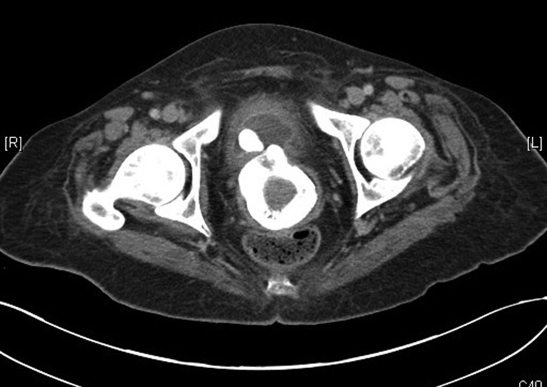
Pelvic CT of patient.
The patient underwent total abdominal hysterectomy and bilateral salpingoophorectomy. During the procedure, a completely calcified aerosol cap was identified in the vagina (figure 2). The cap had been placed in the vagina facing downwards. The foreign object had eroded through the bladder wall and had created a vesicovaginal and right ureterovaginal fistula. The patient had a right ureteric re-implantation and correction of the vesicovaginal fistula.

Surgical specimen.
When interviewed further, she denied any history of sexual or physical abuse. She did not wish to disclose why she had inserted the cap.
Vesicovaginal fistulae secondary to foreign bodies have been reported previously.1 The images highlight the diagnostic challenge we faced because of incomplete history and significant morbidity associated with foreign vaginal bodies.
Learning points
-
Vaginal foreign bodies may have significant sequelae, including acute life-threatening renal failure and fistula formation.
-
History alone may not always highlight the aetiology of symptoms in these sensitive cases.
Footnotes
-
Competing interests None.
-
Patient consent Obtained.