Article Text

Statistics from Altmetric.com
Description
A 26-year-old-man of Indian descent was referred to our rheumatology unit with 6 months of lower-back pain. He had lived in the UK for 5 years, travelling intermittently to holiday in India. His general practitioner detected a C-reactive protein of 55 mg/l and erythrocyte sedimentation rate of 95 mm/h and referred with a possible ‘spondylo-arthritis’.
The patient gave a history of back pain with stiffness throughout the day. He admitted to fever, sweats and significant weight loss but denied having a cough.
On examination, he had slight limitation of lumbar-spine-flexion with no focal spinal-tenderness or clinical-sacroilitis. Straight-leg-raise was 80° bilaterally. Resisted hip-flexion caused exacerbation of lower-back pain. Power and knee-jerks were normal but ankle-jerks were reduced.
Chest-radiograph (figure 1) was normal and lumbar-spine x-ray (figure 2) revealed a fifth lumbar-spine pars-defect only. Renal, liver and bone profile was normal. Three early-morning-urine samples, blood cultures and HIV-serology were negative.

Chest radiograph was normal with no focal parenchymal lung pathology.
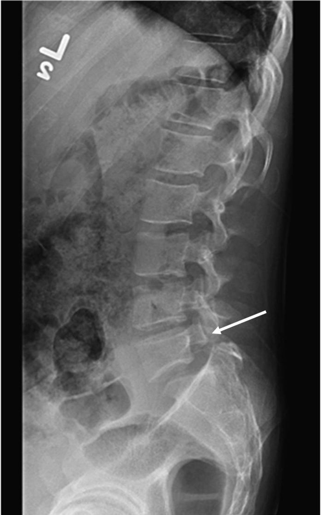
Lumbar spine x-ray demonstrating a fifth lumbar spine pars defect (white arrow).
Fevers, sweats, weight loss and reduced ankle-jerks are considered atypical for a spondylo-arthritis. Malignancy or indolent spinal infection form important differential diagnoses. An urgent MRI-spine (figure 3) revealed multiple spinal-lesions and a para-spinal-abscess eroding the sacrum. Subsequent CT demonstrated a left sixth-rib-lesion, the biopsy and histology of which revealed non-caseous-granulomas and negative acid-fast-bacilli stain. Culture of this specimen yielded Mycobacterium tuberculosis after 4 weeks.

Sagittal MRI view of (A) thoraco-lumbar spine, (B) lumbo-sacral spine, demonstrating contiguous and non-contiguous spinal lesions as well as a 5.8×3.7 cm presacral abscess (white arrows).
About 23% of patients with spinal-tuberculosis (TB) may have normal spinal-plain-radiographs.1 Our case highlights the need for early spinal MRI in patients with suspected spinal TB, even when plain radiographs appear normal. TB-spondylitis frequently involves multiple-adjacent spinal-vertebrae; non-contiguous vertebral involvement, as seen here, is relatively uncommon.2 Confusion with metastatic malignancy is possible; hence a tissue diagnosis is still essential.2
Learning points
-
In suspected cases of spinal tuberculosis (TB), early spinal imaging with MR is required, even if plain radiographs appear normal.
-
A tissue diagnosis should be obtained whenever possible to exclude other pathologies (malignancy/infection) and to determine sensitivity to antituberculous treatment.
-
Non-contiguous TB spondylitis is relatively uncommon but can be present even when plain radiographs appear normal, thus emphasising the need to MRI the whole spine.
Footnotes
-
Competing interests None.
-
Patient consent Obtained.