Article Text

Statistics from Altmetric.com
Description
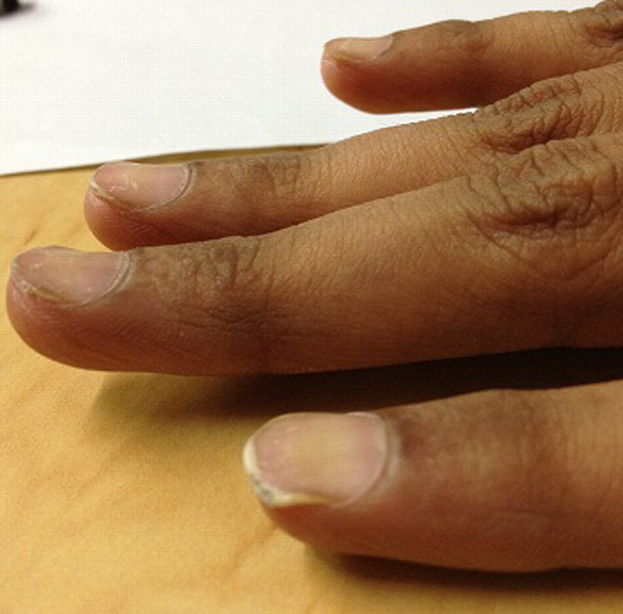
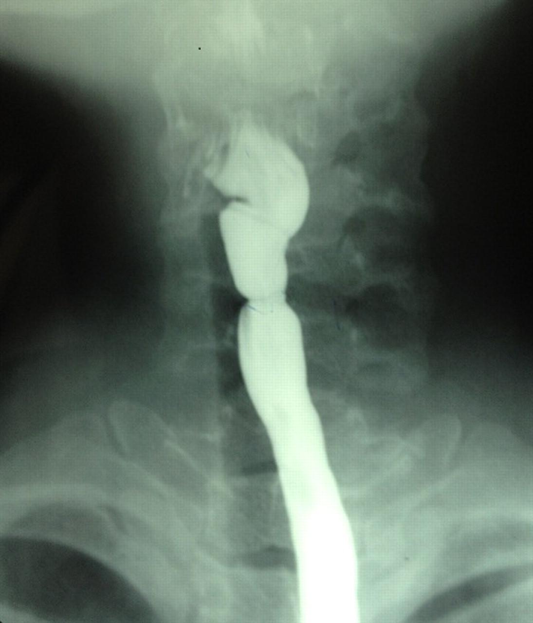
A 27-year-old woman presented to the medicine outpatient clinic with easy fatiguability and breathlessness on exertion since 2 months. She also complained of sore oral mucosa and was unable to swallow large morsels of food. Physical examination revealed angular cheilitis and pigmentation of the oral mucosa, severe pallor, koilonychias (figure 1), large volume pulse and a haemic murmur in the left parasternal area. Investigations revealed hypochromic microcytic anaemia with haemoglobin of 5.6 g%. Serum iron studies, showed an increased total iron binding capacity (TIBC) of 512 µg/l (250–370 µg/l) and low transferring saturation 10% (15–20%) suggesting iron-deficiency anaemia. Barium swallow and upper gastrointestinal endoscopy was done to assess the cause of dysphagia. Barium swallow showed upper oesophageal webs (figure 2). On upper gastrointestinal endoscopy, post cricoids oesophageal web was seen and the endoscope could not be passed across the cricoid level (figure 3). Fulfilling the classical triad of dysphagia, iron-deficiency anaemia and upper oesophageal webs, Plummer-Vinson syndrome (or Paterson-Brown Kelly syndrome, sideropenic dysphagia) was diagnosed. In subsequent endoscopy, the webs were dilated with considerable resultant improvement in her dysphagia. Obvious ulcerated heterotopic gastric mucosa patches were ruled out. Iron supplementation was initiated and the patient was counselled regarding the need for surveillance for the development of an upper gastrointestinal malignancy.

Severe koilonychias.

Upper oesophageal webs seen on barium swallow.

Upper gastrointestinal endoscopy showing oesophageal web.
The pathogenesis of Plummer-Vinson syndrome remains speculative. Iron deficiency has been implicated in the pathogenesis of oesophageal webs and dysphagia in predisposed individuals. The depletion of iron-dependent oxidative enzymes may produce myasthenic changes in muscles involved in the swallowing mechanism, atrophy of the oesophageal mucosa, and formation of webs as epithelial complications. The improvement in dysphagia after iron therapy provides evidence for an association between iron deficiency and postcricoid dysphagia.1 Moreover, the decline in Plummer-Vinson syndrome seems to parallel an improvement in nutritional status, including iron supplementation. Post-cricoid web or Plummer-Vinson syndrome has been identified as a risk factor for the development of upper gastrointestinal tract malignancy.2
Learning points
Plummer-Vinson syndrome classically presents as a triad of iron-deficiency anaemia, postcricoid dysphagia and upper esophageal webs.
Treatment of the iron deficiency frequently relieves dysphagia though some patients may need endoscopic web dilatation.
Patients must be screened regularly for any development of upper gastrointestinal malignancy.
Footnotes
Competing interests None.
Patient consent Obtained.