Article Text

Statistics from Altmetric.com
Description
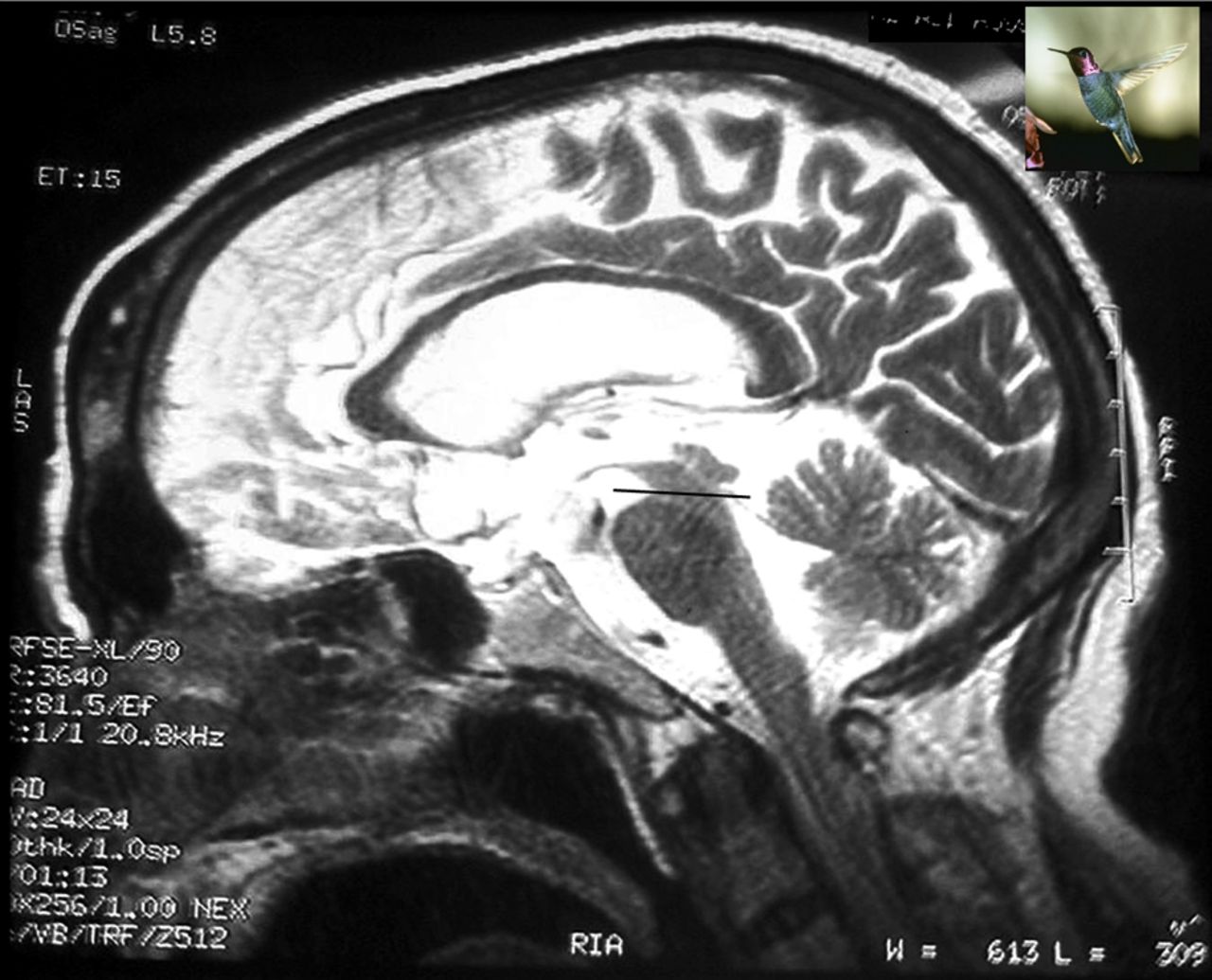
Progressive supranuclear palsy (PSP), previously known as Steele-Richardson-Olszweski syndrome, is an atypical parkinsonian syndrome with a prevalence of ∼5/100 000. It is an important differential diagnosis of more common idiopathic Parkinson's disease (iPD), where clinical differentiation is not straightforward and characteristic neuroimaging often yields a diagnostic clue. We describe a case of 75-year-old man with a history of slowness of activities and recurrent fall while walking, which was insidious in onset and gradually progressive for last 2 years. There was no history of tremor, stooping postures, urinary incontinence or hallucination. On examination supranuclear vertical gaze palsy with slow horizontal saccades, axial rigidity and bradykinesia were present. He had a slow gait with lack of associated movements and tendency to fall backwards. He had positive ‘dirty tie sign’ while eating. The patient was diagnosed as a probable case of PSP and MRI of the brain was done. Mid-sagittal T2-weighted MRI of the brain revealed characteristic selective atrophy of midbrain tegmentum (mesencephalon), with relatively preserved pons, giving an appearance of the head and body, respectively, of a hummingbird (figure 1). This is known as ‘The Hummingbird sign’ or ‘The King Penguin sign’.1 The axial T2 MRI image shows a reduction in the anterior–posterior diameter of midbrain with thinning of cerebral peduncle giving appearance of ‘morning glory sign’ or the ‘Mickey mouse sign’ (figure 2). The hummingbird sign is reported to have a sensitivity of nearly 100%.1

T2-weighted MRI image of the brain showing the selective atrophy of midbrain with preservation of pons (divided by the black line). The atrophy of the midbrain tegmentum results in the concavity forming the silhouette of the head of the ‘Hummingbird’ or the ‘King Penguin’ (inset). This feature is called the hummingbird sign.

An axial T2-weighted image shows the atrophy of the midbrain tegmentum with thinning of cerebral peduncles resulting in the concavity at the lateral margin of the midbrain referred to as the ‘Mickey mouse sign,’ again characteristic of progressive supranuclear palsy.
Image quiz
1. This 75-year-old man had recurrent episodes of fall with slowness of activities and vertical gaze palsy. This is his MR image of brain (figure 1). What is the likely pathology evident from this image?
-
Atrophy of pons with preservation of midbrain.
-
Atrophy of both pons and midbrain.
-
Atrophy of midbrain with preservation of pons.
-
Atrophy of whole of brain stem.
-
Atrophy of fronto-temporal region.
2. A 75-year-old man presented with history of postural instability associated with recurrent episodes of fall and his clinical evaluation revealed vertical gaze palsy, stiffness, preserved occulocephalic reflex with positive ‘dirty tie sign’. Which one of the following radiological signs is expected in the MRI of brain in this patient?
-
‘Hot-cross-bun sign’.
-
‘Hummingbird sign’.
-
‘Giant Panda sign’.
-
‘Knife Blade atrophy’.
Patients with iPD, multisystem atrophy (MSA), corticobasal degeneration do not have midbrain atrophy, thus making this sign important in differentiating these conditions from PSP. This sign also suggests rostral interstitial nucleus of medial longitudinal fasciculus involvement in PSP.2 MR parkinsonism index, defined as the ratio of midbrain area to pons area, is another useful tool in the diagnosis of PSP patients.3 The objective of this article is to highlight the importance of neuroimaging in working up of the differential diagnosis of closely related parkinsonian disorders such as iPD, MSA and PSP.
Image quiz answers
1. (c) Atrophy of midbrain with preservation of pons.
The diagnosis is progressive supranuclear palsy and the mid-sagittal image shows selective atrophy of midbrain with preservation of pons.
2. (b) ‘Hummingbird sign’.
The clinical picture suggests it to be a case of progressive supranuclear palsy (PSP). The T2-weighted mid-sagittal MRI (figure 1) of the brain of this patient shows selective atrophy of midbrain with preservation of pons resulting in the silhouette of the ‘hummingbird’ or the ‘King Penguin’ found characteristically in PSP. ‘Hot-cross-bun sign’ is seen in MRI of brain in multisystem atrophy, ‘Giant Panda sign’ is seen in Wilson's disease and ‘Knife Blade atrophy’ is seen in fronto-temporal lobe degeneration.
Learning points
-
Progressive supranuclear palsy (PSP) is a so-called taupathy with a very low prevalence which has many clinical features overlapping with the more common classical idiopathic Parkinson's disease and differentiation on clinical grounds is difficult.
-
Significant midbrain atrophy with no pons atrophy has been referred to as ‘the Hummingbird sign’ or the ‘Penguin Sign’. This sign is quite useful in differentiating PSP from idiopathic Parkinson's disease and multisystem atrophy. Other imaging findings in PSP that may be seen are elevated apparent diffusion coefficient in the putamen, globus pallidus and caudate nucleus.
-
Abnormal superior profile of midbrain, maximal diameter of midbrain in mid-sagittal plane and MR parkinsonism index are some of the useful keys derived from MRI of the brain that helps in establishing the diagnosis of PSP.
Footnotes
-
Dr. Subhasis Maitra, MD, Assistant Professor, department of General Medicine for his clinical guidance in this case.
-
Competing interests None.
-
Patient consent Obtained.