Article Text

Statistics from Altmetric.com
Description
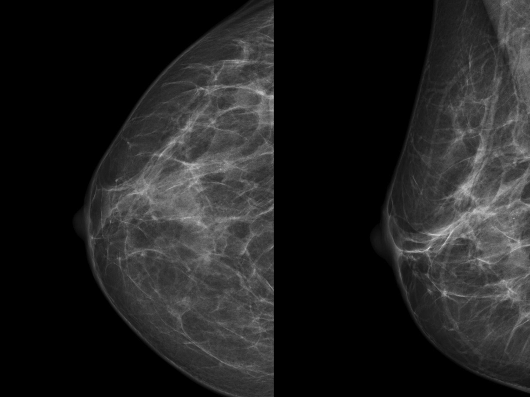
Milk of calcium in the breast consists of calcium deposits within microcysts, and it is found in 4–6%1 of all women undergoing mammography. It is important to recognise its characteristic features in order to avoid unnecessary biopsy (figure 1).2

Craniocaudal and medio-lateral oblique mammographic views showing a single cluster of microcalcifications located in the outer quadrants of the right breast.
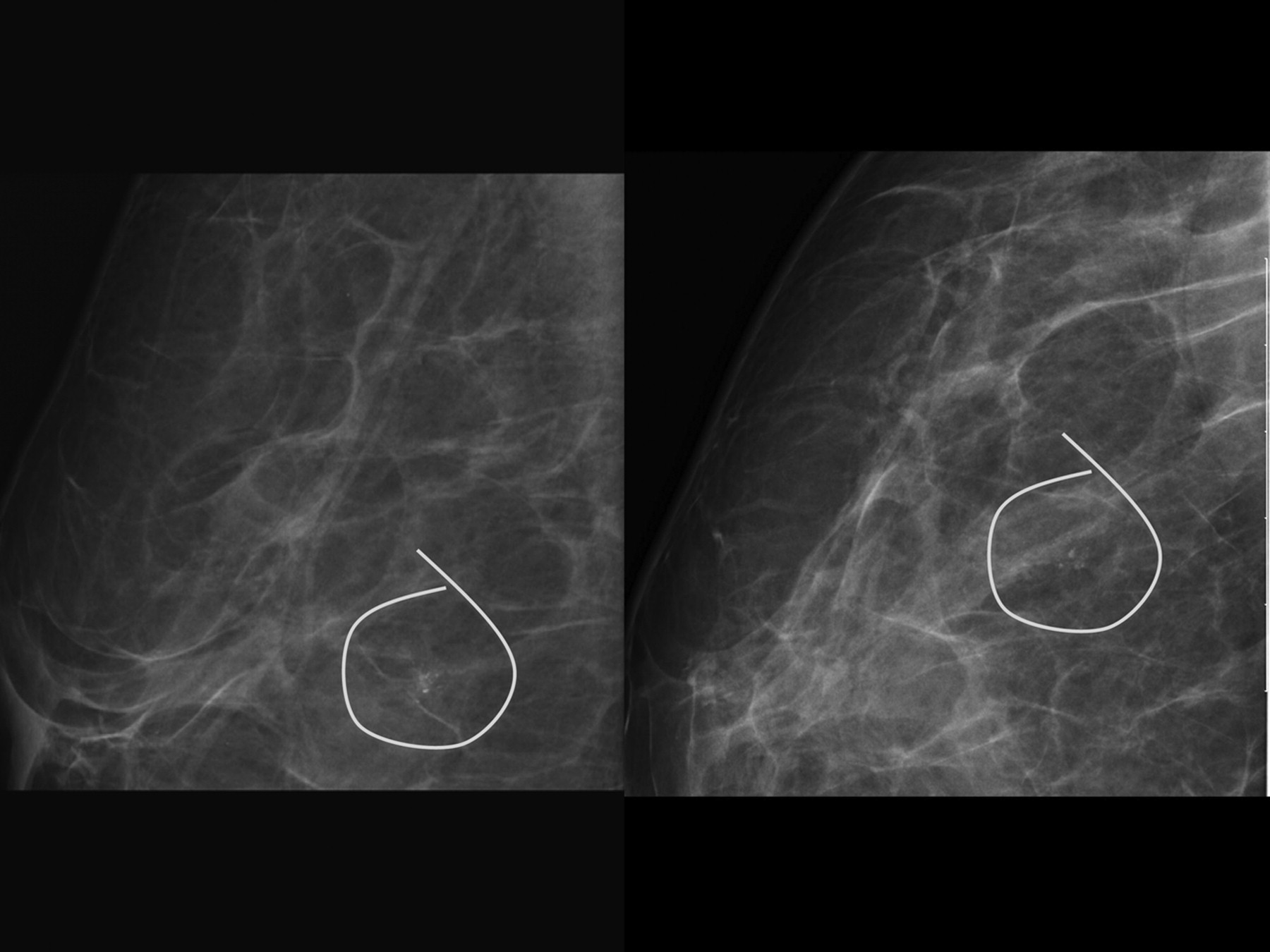
Mammographic appearance is pathognomonic of this benign entity (figure 2).1 Characteristically, calcium particles within cysts produce linear, curved or ‘teacup’-shaped calcifications on medio-lateral oblique or true lateral (TL) views, which clearly change their shape on craniocaudal (CC) views, where the vertical x-ray beam shows smudged calcifications (figure 3).3 This is explained by the deposition of calcium particles at the bottom of the microcysts due to gravity.1

Mammographic views showing the ‘teacup’-shaped microcalcifications on macro true lateral view and smudged calcifications on macro craniocaudal view.

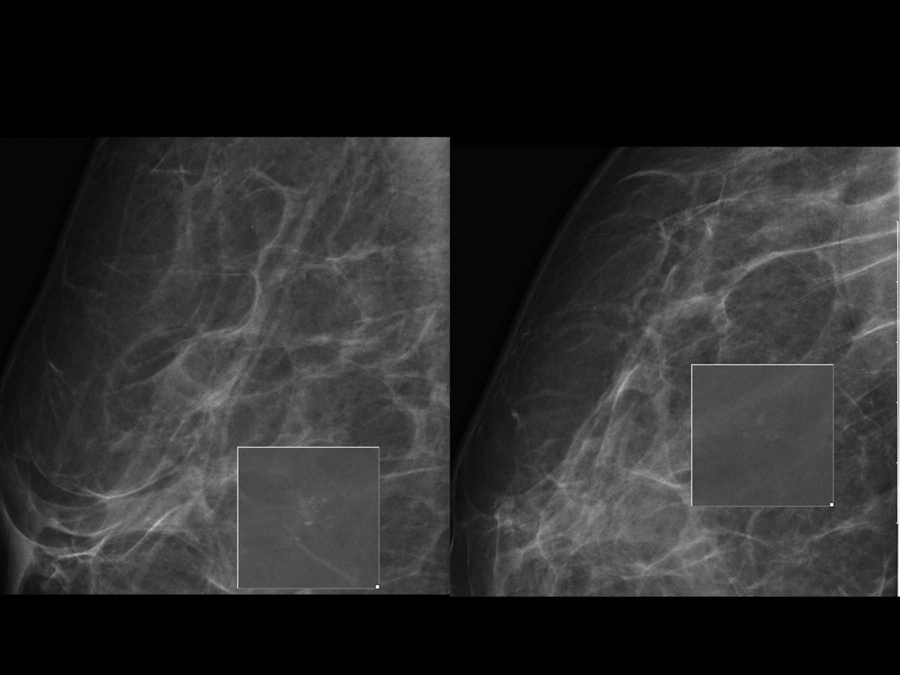
Magnification of mammographic views showing the ‘teacup’-shaped microcalcifications on macrotrue lateral view and smudged calcifications on macro craniocaudal view.
A 44-year-old woman was referred to our breast-imaging department because of the finding of suspicious breast microcalcifications (BI-RADS 4), after voluntary mammography screening in the community. A single cluster of microcalcifications, located in the upper outer quadrant of the right breast on repeat TL and CC magnified views was typical of milk of calcium. The lesion was classified as BI-RADS 2, and the patient was reassured without biopsy.
Conclusion
Mammographic analysis requires expertise and the general principle of biopsying any suspicious or uncertain findings should prevail. Milk of calcium is a relatively common finding, shown to be benign on prolonged follow-up,1 and likely to present at any moderately busy breast-imaging department as a potential cause for unnecessary biopsy. Biopsy should not be performed in such cases, though care should be taken not to miss any suspicious lesions, either in the vicinity or away from the lesion.
Learning points
Suspicious breast microcalcifications should always be biopsied.
“Milk of Calcium” type microcalcifications have been shown to be benign with prolonged follow-up, and shoud not be biopsied.
Benign and suspicious microcalcifications can occur simultaneously, and the latter should be biopsied.
Footnotes
Competing interests None.
Patient consent Obtained.