Article Text

Summary
In this case of secondary syphilis, pustular lesions progressed rapidly to painful ulcerative lesions in a patient with early HIV infection. This rapidly progressive form of early syphilis has historically been called lues maligna praecox, a severe form of noduloulcerative secondary syphilis. Serologic tests for syphilis were positive and biopsy showed forms consistent with Treponema pallidum in the lesions. This case demonstrates how HIV infection may affect presentation and diagnosis of secondary syphilis.
Statistics from Altmetric.com
Background
Ulcerative lesions are a rare presentation of syphilis. We initially presented this case at our weekly Infectious Disease Citywide Conference in Houston, and we were met with open scepticism about the lesions being the result of an HIV and syphilis co-infection. Subsequent biopsy results revealed numerous forms consistent with Treponema pallidum in the lesions, and a photographic literature review on syphiloderma ulcerativum from the 1800s (pre-HIV era) supported our case report. We wrote about this unusual presentation of a common disease to remember a forgotten form of syphilis.
Case presentation
A 34-year-old man with newly diagnosed HIV infection presented with 1 month of progressive, painful nodular and ulcerative skin lesions.
Four months before presentation, he had a diffuse, maculopapular rash that spared his palms and soles together with subjective fevers, rhinorrhoea and a sore throat. His primary care physician gave him medications for presumptive chickenpox and, within 1 month, the rash resolved.
One month before admission, he developed pustules, some of which enlarged and ulcerated. They were large and varied in depth and circumference along his left mandible, right axilla, right chest wall and left perineal region. His primary care physician prescribed clindamycin.
Two weeks before admission, the patient presented to the emergency room because his ulcers were worse and now had become painful. He reported no fevers, chills, night sweats, weight loss, recent illness or sick contacts. When the pain became unbearable, he re-presented to emergency room and was admitted for further evaluation. He reported a negative HIV test and negative rapid plasma reagin (RPR) approximately 12 months before admission.
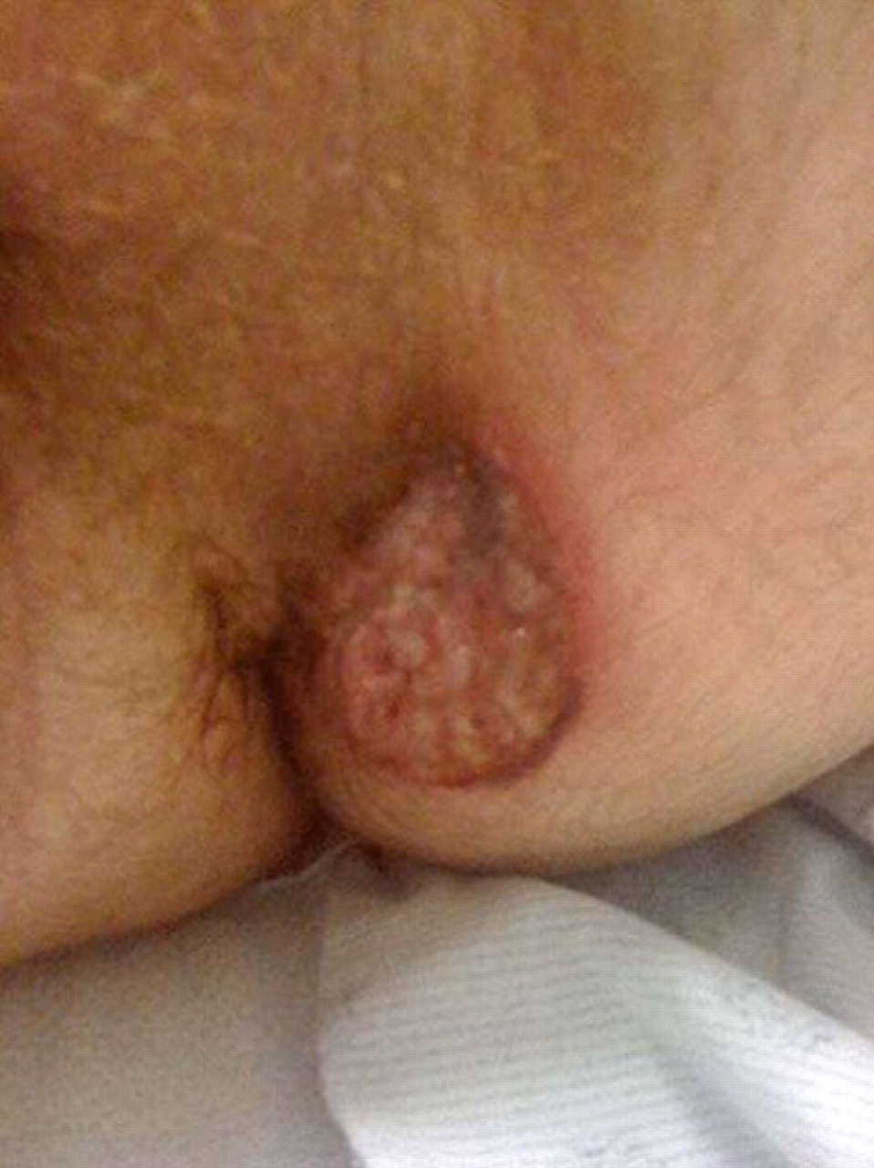
The patient had normal vital signs. He was in mild distress. The skin had scattered pustules and approximately 10 ulcerative lesions ranging from 1×1 cm to 3×5 cm were present on his trunk, face, arms, axilla and gluteal fold. The largest one extended to muscle while the smallest one did not extend beyond subcutaneous tissue. The borders were well demarcated and erythematous with associated crusting (figures 1 and 2). He had no genital lesions, but a rectal fissure was present. Neurologic examination revealed intact cranial nerves, normal sensation and a normal gait.

The face (left mandible) is a typical location for ulcerative syphilides.

The ulcerative syphilide at the gluteal fold appears in the distribution of condyloma lata (kissing lesions).
Investigations
His complete blood count was normal as were other routine laboratory tests. RPR was positive at 1:64 and Treponema pallidum particle agglutination assay was positive. HIV enzyme immunoassay and Western blot were positive. HIV-1 viral load was 930 000, and CD4 count was 408 cells/mm3 with 17% CD4 cells.
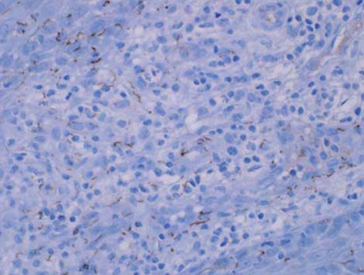
Wound cultures showed normal skin flora. Skin punch biopsy showed necroinflammatory debris lining the ulcer base, with a dense mixed inflammatory infiltrate of lymphocytes, histiocytes, plasma cells, neutrophils and eosinophils permeating the superficial and deep dermis. The vascular plexus contained reactive and swollen endothelial cells with mild proliferation (figures 3 and 4). Fite’s acid fast, Gomori methenamine silver and gram stains were negative. Herpes simplex virus was not detected. An immunohistochemical stain directed against spirochetes demonstrated numerous corkscrew-shaped microorganisms (figure 5).

Low power view shows subtotal epidermal ulceration and a dense mixed inflammatory infiltrate within the superficial and deep dermis.

High power view illustrates reactive endothelial cells and scattered plasma cells, eosinophils and neutrophils within a dense lymphohistiocytic inflammatory infiltrate.

Immunohistochemical stain directed against spirochetes reveals numerous corkscrew-shaped microorganisms, consistent with the morphology of T pallidum.
Treatment
The patient was treated with three doses of 2.4 × 10⁶ units of benzathine penicillin at weekly intervals. Close clinical monitoring confirmed that he did not have a Jarisch–Herxheimer (JHR) reaction.
Outcome and follow-up
One month after the first dose of penicillin, his ulcers had formed scars that completely healed, and his pain had fully resolved.
Discussion
This patient with HIV infection of recent onset was diagnosed as having lues maligna praecox – a rare but severe form of secondary syphilis. Numerous pustular lesions appeared. Over a period of several weeks, the lesions progressed to extremely painful ulcerations. In the preantibiotic era, such lesions were called ulcerative syphilides.1 2
Although lues maligna was described well before the HIV pandemic,3 more cases of lues maligna have been described in people with HIV infection.4,–,7 Persons living with HIV/AIDS are 60 times more likely to present with this form of syphilis.8 Immunologic events that facilitate the development of lues maligna are unknown, but it is reasonable to postulate that the loss of helper T cells is responsible. CD4 cell counts have been low in those few previous cases in which they have been reported. These findings suggest that people with acute HIV infection may be an at-risk population.
The lesions of lues maligna may present a diagnostic dilemma in the HIV population. Physicians rely on the following diagnostic criteria developed in the pre-HIV syphilis era: (1) compatible gross and microscopic morphology; (2) a high titre serologic test for syphilis; (3) a severe JHR; and (4) dramatic response to antibiotic therapy.9 Our patient met only two of these four diagnostic criteria for lues maligna: comparable gross appearance and a dramatic response to penicillin. Of course, the immunohistologic stains for treponemes were not available when the original criteria were developed.
We diagnosed secondary syphilis instead of tertiary syphilis based on the biopsy findings. The lesion showed a mixed inflammatory infiltrate with epidermal ulceration rather than the expected plasma cell predominant dermatitis.4 Treponemes are rarely detected in syphilitic gummas, although in one case, treponemes were detected using indirect immunofluoresence.10 In our case, the presence of numerous treponemes on immunohistochemical stain diagnosed secondary syphilis with certainty and excluded the possibility of a gumma.
Learning points
▶ Lues maligna praecox is a rare but severe form of secondary syphilis in which pustular lesions progress rapidly to painful ulcerative lesions.
▶ HIV infection may affect presentation and diagnosis of secondary syphilis.
▶ Persons living with HIV/AIDS are 60 times more likely to present with this form of syphilis.
▶ The presence of numerous treponemes on immunohistochemical stain can diagnose secondary syphilis with certainty and exclude the possibility of a gumma.
Acknowledgments
The authors would like to thank the patient for sharing his story.
Footnotes
-
Competing interests None.
-
Patient consent Obtained.