Article Text

Statistics from Altmetric.com
Description
Hydatid disease is caused by infection with the tapeworm Echinococcus granulosus. At 5–10%, the prevalence rate in Kenya’s Turkana region is among the highest worldwide.1 Infection involves cyst formation in many sites of the body, with cardiac hydatid disease being rare (1% of all hydatid infestations) but potentially fatal.2
We report the case of a man in his late 30s, referred by one of the authors for management of cardiac hydatid disease. He had presented to his local clinic with exertional angina, occasional haemoptysis and shortness of breath. An interventricular cyst had been demonstrated on ultrasound, and the patient was transported by flying doctor to our charity-based Kenyan heart surgery centre.
Clinically, we found the man to be healthy, with cardiac and respiratory examination being unremarkable. Hydatidosis was diagnosed on echocardiography, with a 4.9×4.7 cm immobile cyst located in the interventricular septum (figure 1). ECG revealed ST segment depression and T wave inversion in leads V5 and V6. Chest radiography and ultrasound demonstrated two cysts within the right pleural cavity. Abdominal ultrasound was normal and CT unavailable.

(A and B) Echocardiogram showing interventricular cyst.
On cardiopulmonary bypass, the anterior descending coronary artery was found to be crossing anteriorly to the cyst, explaining the unusual presentation of the cyst with angina. Only around four cases of cardiac hydatid disease presenting with angina have been reported, with only one due to an interventricular cyst.3
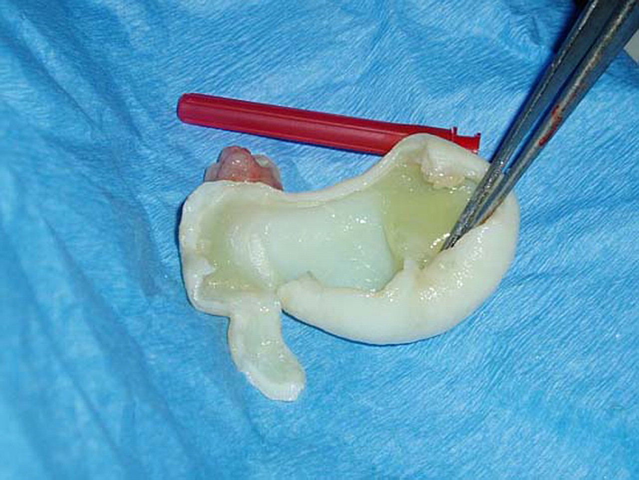
Cystopericystectomy was performed with a fine needle, aspirating the contents (15 ml) before removing the pericyst (figure 2). The patient later underwent a thoracotomy to remove the pulmonary cysts, making a satisfactory recovery and returning to normal activities.

The pericyst after aspiration of its contents.
Acknowledgments
The authors acknowledge the work undertaken by the team at the Kenyatta National Hospital in facilitating this treatment.
Footnotes
-
Competing interests None.
-
Patient consent Not obtained.