Article Text

Statistics from Altmetric.com
Description
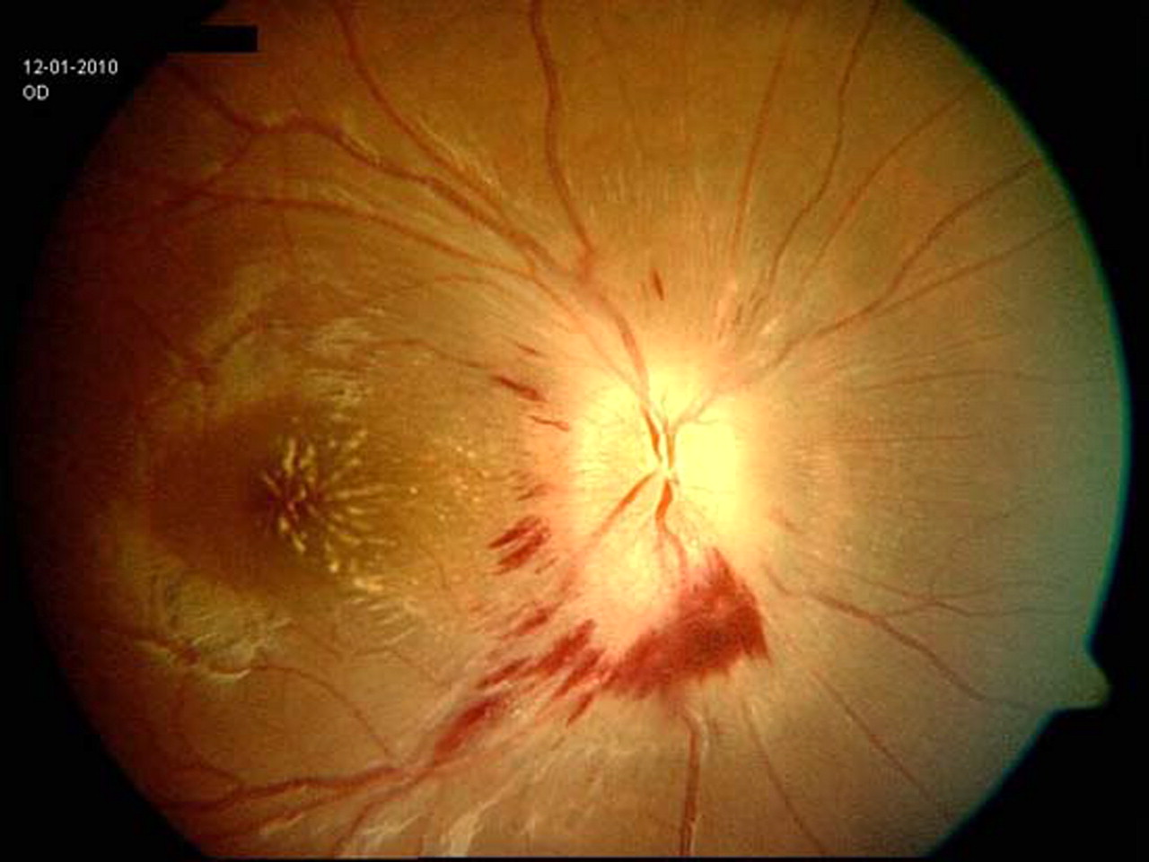
A 20-year-old girl with acute lymphoblastic leukaemia in second remission for 4 years, presented with blurring of vision and binocular diplopia for 2 months duration. Examination revealed isolated left 6th cranial nerve palsy. Fundus showed swollen both optic disc and fan-shaped macular star suggestive of papilloedema. CT scan and MRI of brain revealed no space occupying lesion or hydrocephalus. However the presence of peripapillary yellowish infiltrates raised suspicion of leukaemic infiltrates (figures 1 and 2). Lumbar puncture revealed numerous blast cells. Bone marrow biopsy also showed frank relapse with precursor B cells acute lymphoblastic leukaemia. Leukaemic optic disc infiltration is rare and may mimic papilloedema.1 Presence of yellowish peripapillary infiltrates2 should alert attending physician to proceed with lumbar puncture to confirm the diagnosis of a relapse. This patient underwent systemic and intrathecal chemotherapy.

Right fundus image shows peripapillary yellowish infiltrates and haemorrhages.

Left fundus image shows peripapillary yellowish infiltrates and haemorrhages.
Footnotes
-
Competing interests None.
-
Patient consent Obtained.