Article Text

Statistics from Altmetric.com
Description
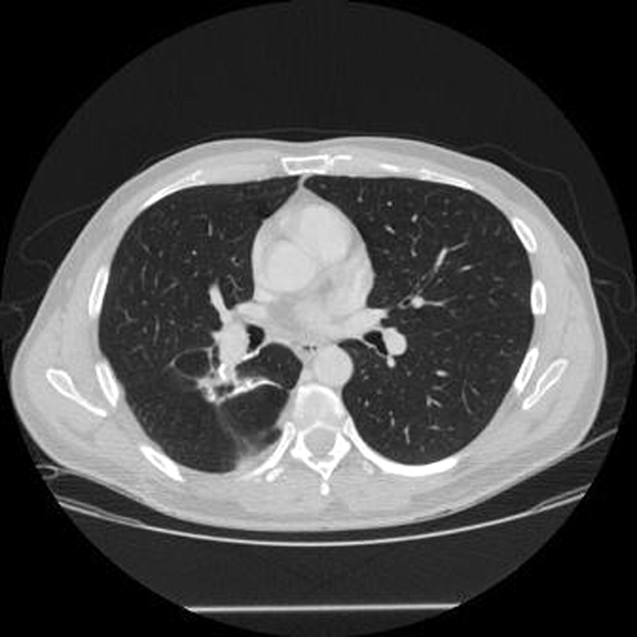
A 46-year-old male has a recent history of rectal adenocarcinoma ypT3N1M0G2, treated by neoadjuvant radio-chemotherapy, low anterior rectal resection and adjuvant chemotherapy. Before surgery a thoraco-abdominal CT showed no other pathological lesion. The patient presented free of symptoms 6 months after surgery for follow-up visit, had no fever and normal values at routine laboratory evaluation. The thoraco-abdominal CT identified a cystic lesion with thin wall in the apical segment of the right inferior lobe of 2.5 cm diameter, not seen at previous radiological assessment (figure 1), with no thoracic lymphadenopathy and without signs of abdominal disease; it was considered that colonoscopy was not necessary at the time. The cystic lesion was interpreted initially of unknown origin, the patient presenting neither inflammatory signs nor respiratory symptoms. His metastatic origin could not be ruled out at the time, the carcinoembryonic antigen being normal. The patient was followed strictly and 6 months later a new CT (figure 2) showed growth in size of the lesion that appeared with a thicker wall, both features sustaining the metastatic probability. There was no liver metastasis at CT and colonoscopy at 1 year after surgery found no local recurrence. Cardiopulmonary tests were normal without contraindication of thoracic surgery. The patient had right thoracotomy and wedge resection of the pulmonary lesion. The pathological examination revealed a metastasis of adenocarcinoma of colorectal type, without infiltration of the pleura and with 50% viable cells, negatives for cytokeratin 7 and thyroid transcription factor-1 at immunohistochemistry. The patient had another regimen of adjuvant chemotherapy (six cycles) by 5-fluorouracil, leucovorin and avastin, even if the treatment was already completed previously following the international consensus for the stage of the colorectal cancer. At 1 year follow-up, the patient was free of symptoms and without radiological evidence of metastatic disease (figure 3). Cystic lung lesions can be caused by a diverse array of pathologic processes. Evaluating a patient with such lung lesions is helpful to distinguish cysts from cavities and to categorise focal or diffuse distribution. Cysts are defined as air-containing spaces surrounded by a thin wall (4 mm or less) while cavities are air-containing spaces with walls that are at least 5 mm thick.1 One of the most important distinctions in the differential diagnosis of cystic or cavitary lung lesions is the distinction between malignant and benign aetiologies. Primary lung cancer is a common disease, cavitation being detected by CT for up to 22% of primary lung cancers but for near 11% by plain thoracic radiography. Cavitation more frequently occurs in squamous cell carcinomas than in other histological type. Furthermore, the presence of cavitation in a lung tumour has been associated with a worse prognosis. Other primary tumours in the lung, such as lymphoma and Kaposi’s sarcoma, may also present with cavitary lesions, particularly among persons infected with human immunodeficiency virus.2 Metastatic lung disease may also have a cavity or cystic appearance, but less frequently than in primary lung cancers: between metastatic tumours less than 5% were found cavitary by plain radiography. Metastatic tumours of squamous cell origin are more likely to cavitate than tumours of other origins, suggesting a common pathogenesis for cavitation among these tumours.2 3 Infections caused by commonly encountered bacteria may cause pulmonary cavities by one of two mechanisms. First, organisms may enter the respiratory cavity via the upper airway and cause either a necrotising pneumonia or lung abscess. Alternatively, organisms may enter the lung via the bloodstream, often in association with fibrin and platelets as septic pulmonary emboli.2 Complicating the diagnostic evaluation of cavitary lung lesions there are several reports that have revealed the coexistence of pulmonary malignancy and an infectious pathogen. Non-infectious inflammatory processes associated with cystic pulmonary lesions include various vasculitides such as Wegener granulomatosis and rheumatoid nodules. Pulmonary embolism can also causes cystic lesions especially in immunosuppressed patients, even if infarction is found in less than 15% of cases, and only about 5% of infarctions cavitate.1 In our case, the patient had previous oncologic history and few risk factors for a primary lung tumour. The infectious origin was not sustained by biological tests and in spite of the rarer presentation; the metastatic origin was our first diagnosis considering the patient’s situation. In conclusion, even the spectrum of pathological processes associated with pulmonary cavities and cysts is extremely large, narrowing that the differential diagnosis can be facilitated by a careful review of the patient’s history and imaging data, making the clinical context as the most useful diagnostic tool.

Cystic pulmonary lesion in the right inferior lobe appeared 6 months after rectal surgery.

The cystic lesion has grown in size in the following 6 months, sustaining the metastatic origin.
CT of the thorax at 1 year after lung metastasectomy showing scars and staples at the operated area in the right lung.
Footnotes
-
Competing interests None.
-
Patient consent Obtained.