Article Text

Statistics from Altmetric.com
Description
A 17-year-old female patient was admitted to our hospital with large skin defects around her ileostoma since 6 months. Several years before, she underwent a colectomy due to colitis ulcerosa and an ileostoma was created. A sharply demarcated, deep peristomal ulcer was observed (figure 1). Despite intravenous antibiotics, the ulcer worsened and caused pain. Bloodcultures remained negative. The clinical diagnosis pyoderma gangrenosum (PG) was made, but skin biopsy was not performed because of the typical appearance. Treatment with 60 mg oral prednisone was initiated and the ulcer rapidly improved (figure 2). PG is a rare, non-infectious, ulcerative skin disorder. Approximately 50% of the cases are associated with a variety of systemic diseases, such as arthritis, leukaemia or inflammatory bowel disease. Of the latter 0.6–5% develop PG. The pathogenesis is poorly understood. It is thought to be an immune dysregulation disorder with extensive neutrophil inflammation of the skin with vasculitis-like features. It most commonly occurs in women. Only 4% of all patients are children.1 Histology is aspecific. Diagnosis is made by clinical appearance and non-improvement after conservative or antibiotic treatment and is therefore often delayed. PG can occur all over the body.1 2 It often develops at sites of trauma such as biopsy and surgical scars. The first choice of treatment is systemic steroids combined with conservative wound care. However, treatment of the underlying disease is necessary.1 PG may heal after colectomy, although, like in our case, it has been described after colectomy.2 The prognosis remains unpredictable.
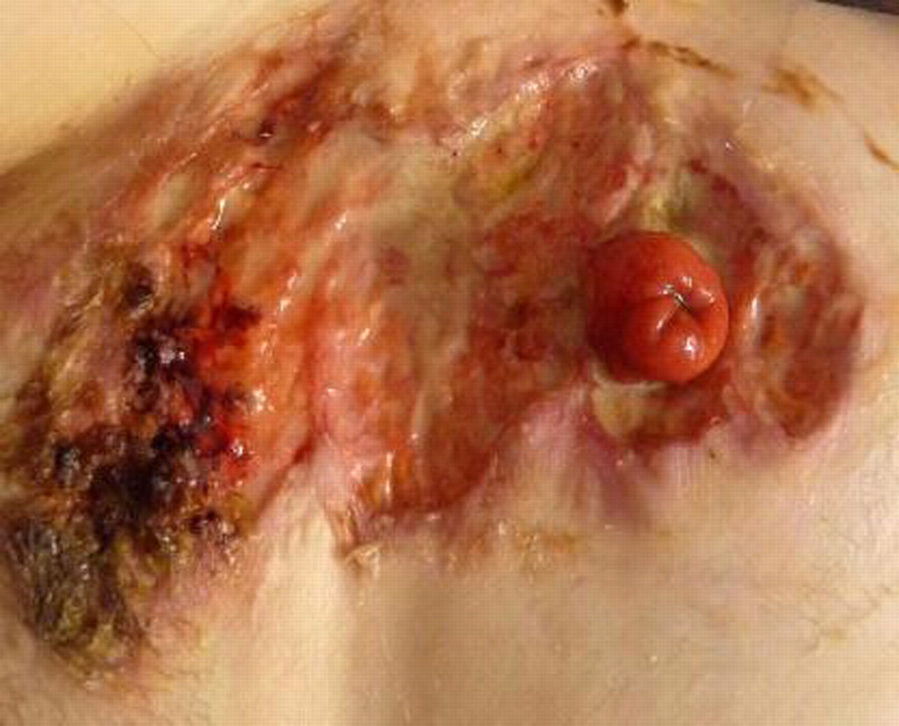
Peristomal lesion with yellow, red and black appearance and elevated borders, partly overhanging the ulcer bed.

Peristomal lesion during treatment.
Footnotes
-
Competing interests None.
-
Patient consent Obtained.