Article Text

Summary
An 81-year-old Caucasian lady with permanent atrial fibrillation (AF) was admitted with palpitations and fast AF. She took bisoprolol and warfarin with subtherapeutic international normalised ratio. Rate control of AF was successful. Around 36 h later, she developed abdominal pain and vomiting. A caecal volvulus was diagnosed on CT. She underwent emergency laparotomy. Intraoperatively, an obstructing tumour was found in the colonic hepatic flexure. It was deemed inoperable. The caecal volvulus was decompressed and an ileo-transverse colon bypass was performed. She made a good recovery and her fast AF settled postoperatively. She was seen by the oncology team and was discharged with palliative care support with no further exacerbation of AF.
Statistics from Altmetric.com
Background
Atrial fibrillation – an age old disease
Atrial fibrillation (AF) and atrial flutter (AFL) are the commonest narrow complex arrhythmias. They are known to complicate underlying malignancies.1 Liu and Pusalkar described a case of new onset AFL in a patient with glioblastoma multiforme. However, the authors concluded that the association was likely to be coincidental. AF is the most prevalent arrhythmia which affects 1–2% of the population.2 It is age dependent, affecting less than 0.5% of people in their fourth decade, rising to more than 10% in subjects over 80 years old.2 Despite extensive research into its pathogenesis and management, the incidence and prevalence of AF is increasing.3 According to both the National Institute of Clinical Excellence guidelines (NICE) and the 2006 American Heart Association/American College of Cardiology/European Society of Cardiology guidelines (AHA/ACC/ESC); AF is classified into three groups.3 Paroxysmal AF lasts less than 7 days and terminates spontaneously while persistent AF lasts more than 7 days and may terminate spontaneously or are successfully cardioverted to normal sinus rhythm (NSR).3 Permanent AF is present if the arrhythmia lasts more than 7 days and cannot be cardioverted back into NSR.3 However, these terms are not mutually exclusive. The duration of AF reflects the length of each individual episode as well as the total amount of time a patient has suffered from AF since diagnosis.3 For this reason, a patient who suffers many years from recurrent episodes of AF with spontaneously terminations can be classified as paroxysmal.3
The pathogenesis of AF is complex. Initiation is likely due to a combination of atrial anatomical pathology as well as electromechanical remodelling.3 Atrial fibrosis and atrial muscle loss are present in early AF and can be demonstrated in biopsies. Atrial fibrosis occurs adjacent to normal conducting atrial tissue, providing heterogeneous conduction substrates for arrhythmogenesis.4 Patients with severe atrial fibrosis are less likely to respond to cardioversion than those with mild to moderate fibrosis.3 Atrial dilatation is also implicated in the onset and perpetuation of AF, which may be caused by activation of the renin-angiotensin-aldosterone system (RAAS).3 Propagation of AF worsens any atrial dilatation present at onset, leading to decreased contractility and reduced atrial compliance.3 Interestingly, atrial stretch induced by AF upregulates RAAS and transforming growth factor β-1, contributing to further atrial fibrosis.5 These structural changes are likely to provide the substrate for electrical disturbances.
There are two hypothesised mechanisms for the electrical disturbances in AF: the automatic focus theory and the multiple wavelet theory.3 In the former, there is a single arrhythmogenic focus or multiple foci within a discreet area.3 The commonest region is near the junction between the left atrium and the pulmonary vein (LA-PV), owing to the shorter refractory period of atrial tissue in the PV.6 Indeed patients with AF have shorter refractory periods within their PV compared to controls.6 Rapid-firing from foci within the PV may lead to micro-reentry, exacerbated by shorter refractory periods.7 Catheter ablation near the LA-PV junction has led to successful termination of AF in selected patients.3
The multiple wavelet theory, first described by Moe and colleague, refers to chaotic firing of multiple wavefronts within the atria from several foci.8 The number of wavefronts is increased by a large atrial mass, reduced conduction velocities and shortened refractory periods.3 This theory explains why atrial fibrosis (reduced conduction velocity) and atrial dilatation (large atrial mass) predispose to AF.3 There also appears to be a gradual, albeit controversial, progression from paroxysmal to persistent AF culminating finally to permanent AF.3
Caecal volvulus – a rare surgical phenomenon in practice?
Caecal volvulus is defined as torsion of the caecum on its mesentery, leading to nearly 1% of all cases of bowel obstruction.9 Causes include abnormal colonic fixation to the peritoneum to allow a free segment to twist on itself.9 In addition, abdominal masses and adhesions can serve as rotatory fulcrums.9 The most serious complication is closed-loop obstruction leading to bowel necrosis and death if prompt surgical management is delayed.10 Presentations are usually those of bowel obstruction: vomiting, constipation, colicky abdominal pain and distension.11 Classically, caecal volvulus can be diagnosed on abdominal radiographs as isolated distended bowel loop resembling a large ‘coffee bean’.12 This loop usually localises to the left side.12 However, this is unpredictable and a CT is required to distinguish between caecal and sigmoid volvulus.9 The definitive management is surgical intervention with laparotomy and detorsion of the volvulus.9 The usual practice is detorsion with an ileocolic resection with ileocolic anastomosis, with no chance of recurrence.9
Case presentation
An 81-year-old Caucasian lady with a 30 years history of permanent AF, type II diabetes mellitus and diverticular disease presented at 3 a.m with sudden onset palpitations which woke her up from sleep. She also had shortness of breath. There was no chest pain, cough, sputum or haemoptysis. She had no urinary symptoms and there was no change in bowel habit. She took warfarin for AF, bisoprolol, metformin. Her diverticular disease was inactive for 7 years. There was no history of ischaemic heart disease, hypertension or cerebrovascular disease. She was a non-smoker with an unremarkable family history. On initial assessment, she was alert and orientated with a tachycardia at 138/min, auricular temperature of 36.6°C, blood pressure 95/42 mm Hg, respiratory rate 22/min and pulse oximetry was 96% on room air. On examination, her pulse was irregularly irregular. Jugular venous pulse was 2 cm. Normal first and second heart sounds were present with no murmurs or added sounds on auscultation. Diffused fine inspiratory crepitations were heard bilaterally throughout the chest. On admission, her abdomen was soft and non-tender and she was neurologically intact.
Her initial bloods were as follows: haemoglobin 14 g/dl, white cell count 6.5×10⁹/l, platelet 249×109, C-reactive-protein 12.7 mg/l, sodium 141 mmol/l, potassium 4.4 mmol/l, urea 7 mmol/l, creatinine 62 mmol/l, corrected calcium 2.47 mmol/l, international normalised ratio (INR) 0.9 (target range 2–3). Her liver function and thyroid function tests were normal. Serum troponin I was <0.032 µg/l on admission and again at 12 h after the onset of palpitations. Arterial blood gas sample showed metabolic acidosis, hypoxia and raised lactate. She was given oxygen therapy. Her ECG showed fast AF with ventricular rate of 138/min (figure 1). A urine dipstick was unremarkable and her chest radiograph showed evidence of pulmonary fibrosis with cardiomegaly despite antero-posterior film (figure 2).

A 12-lead ECG taken on admission showing fast atrial fibrillation with a ventricular rate of approximately 138/min.

Chest radiograph on admission showing cardiomegaly and fibrotic changes.
She was monitored on telemetry. AF strict rate control was attempted with bisoprolol and digoxin which reduced her ventricular rate to between 80 and 110 for the next 48 h. Her subtherapeutic INR was noted and she was given increased doses of warfarin with low-molecular weight heparin cover until therapeutic. A transthoracic echocardiogram showed a right atrial size of 5.5 cm with left ventricular ejection fraction of 50%. There was evidence of trivial mitral regurgitation.
Thirty-six hours after admission, the patient developed sudden onset right iliac fossa pain 3 h after a light lunch with faecolant vomiting. On examination, her abdomen was distended with guarding and tenderness over the right iliac fossa. No rebound tenderness or masses were detected. Bowel sounds were hyperactive. Digital rectal examination was unremarkable. Abdominal radiograph showed right-sided large bowel distension indicative of caecal volvulus (figure 3), which was confirmed with CT abdomen/pelvis (figure 4). The CT scan also showed ischaemic changes surrounding the volvulus and the large bowel wall. She was reviewed by the surgical team and underwent an emergency laparotomy. Intraoperatively, an obstructing tumour was found in the hepatic flexure inseparable from the duodenum and pancreas. It was deemed inoperable. Her caecal volvulus was decompressed via an appendicectomy. An ileo-transverse colon bypass was carried out.
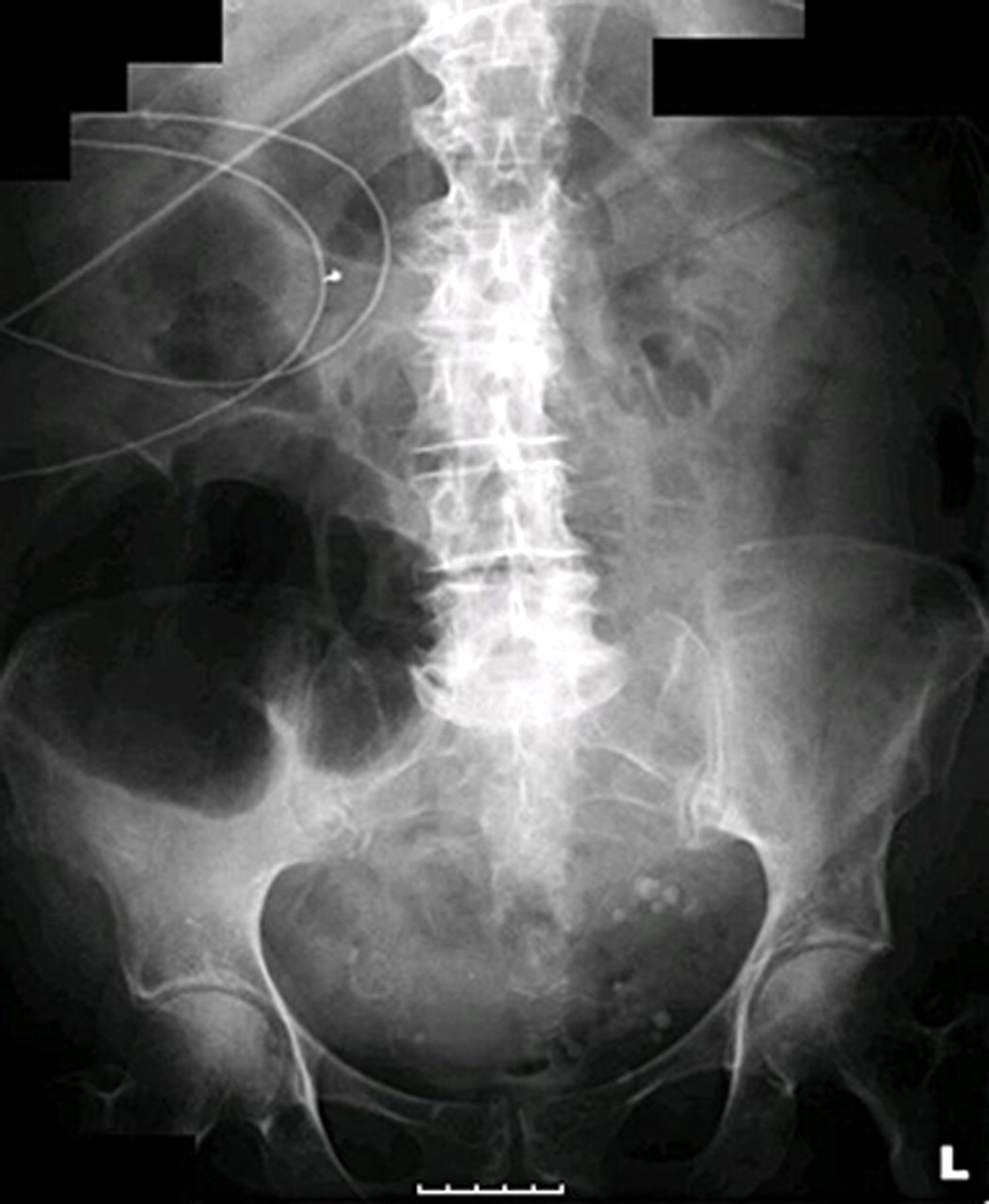
Abdominal radiograph demonstrating a grossly distended loop of bowel, likely a volvulus.

CT demonstrating a caecal volvulus.
Outcome and follow-up
Postoperatively, the patient made a good recovery in the intensive care unit (ITU). On the second day in ITU, her fast AF was well controlled with normal ventricular response below 80/min on digoxin and bisoprolol, which were continued. She was referred to the oncology team for the management of malignancy, which unfortunately never obtained a histological diagnosis since no biopsies were taken intraoperatively. She was later found to have profoundly raised CA19.9 and carcinoembryonic antigen tumour markers. A lengthy discussion took place between the oncology consultant, the patient and her relatives and a joint decision was made for palliative management given her age and co-morbidities. She was discharge a week later with palliative care services and follow-up with the oncology team. She did not have another relapse of fast AF.
Discussion
We described an older lady who presented with fast AF and subsequently developed caecal volvulus most likely secondary to an obstructing tumour in the hepatic flexure. This case demonstrates two interesting principles. First, it gives an account of the challenging task of managing fast AF in older patients. The successful management of AF in the acute setting is sometimes difficult and an evidence based outline is not always available. Second, this case demonstrates the unpredictable nature of clinical practice where two rather contrasting ailments (AF and caecal volvulus) can coexist. This necessitates an open minded approach in cardiology; a reminder of an important clinical lesson.
The NICE management of AF
AF management can be broadly divided into rate or rhythm control. The latter aims to restore NSR. Stroke prevention should be carried out with either intervention.3 According to the NICE guidelines, provided that adequate thromboprophylactic measures are taken, permanent AF should be managed with rate control and paroxysmal AF with rhythm control. Persistent AF can be managed with either rate or rhythm control depending on patient related factors. Rhythm control is preferred in younger, patients with symptoms with first presentation of lone AF or those with congestive heart failure. Rhythm control should also be attempted if AF is secondary to treated precipitants. Rate control is better indicated in sufferers of persistent AF if the patient is older (over 65 years), with coronary artery disease and unsuitable for cardioversion and antiarrhythmic drugs.
Rate control versus rhythm control
Two landmark non-inferiority trials AFFIRM13 (Atrial Fibrillation Follow-up Investigation of Rhythm Control) and RACE14 (Rate Control versus Electrical Cardioversion for Persistent Atrial Fibrillation) have helped to solve the age old conundrum of rate versus rhythm control. The AFFIRM trial enrolled 4060 patients randomised to receive either rate control or rhythm control. The intervention in the latter arm of the study was achieved with antiarrythmic drugs and direct-current cardioversion.13 Stroke prevention with warfarin (target INR 2–3) was offered to all rate controlled subjects and those in the rhythm control group with a minimum period of 4–12 weeks in NSR.13 There was no significant difference in the 5-year mortality between the rate controlled and rhythm controlled arms; 21.3% versus 23.8%, respectively. CI of 0.99 to 1.34 (p=0.08).14 The stroke rates were also comparable among the rate controlled and rhythm controlled patients.13 The RACE trial was a randomised controlled trial of 522 patients comparing rate control and rhythm control. The authors concluded that AF rate control was not inferior to rhythm control in reaching primary end points.14 These end points included cardiovascular death, heart failure hospitalisations, severe bleeding, thromboembolism, pacemaker insertion and severe drug side effects.14 Patients in the rhythm controlled group experienced more drug induced side effects and slightly higher thromboembolic events.14
The target heart rate in rate controlled AF is a controversial entity. Recently, RACE-II (Rate Control Efficacy in permanent AF) trial assessed whether strict rate control (<80/min resting, <110/min moderate exercise) was superior to lenient rate control (<110/min resting).15 Six hundred and fourteen patients with AF were enrolled and randomised to either strict or lenient rate control and followed up for a maximum of 3 years.16 Similar primary end points were used as in earlier AFFIRM13 and RACE14 trials. The investigators concluded that lenient rate control was non-inferior to strict rate control.16 This approach has been adopted by both the ESC17 and the ACC Foundation/AHA and Heart Rhythm Society (ACCF/AHA/HRS).18 The ESC recommends lenient rate control as first line; commencing therapy with resting heart rate of <110/min. Strict rate control is second line once lenient control fails to prevent symptoms or complications such as cardiomyopathy.15 ACCF/AHA/HRS guidelines state that strict rate control has no benefit.17 In contrast, NICE guidelines adopts strict rate control (<90/min resting, <110/min with exercise) in all cases except for recent-onset AF, for which lenient rate control is applied (target <110/min).19 The variations in the guidelines reflect the limited volume of evidence on strict versus lenient rate control.
The patient was likely to be suffering from ‘lone’ AF since there was no history of precipitants such as ischaemic heart disease, vavlular disease, alcoholism or thyrotoxicosis. In all likelihood, the cardiomegaly present on chest radiograph was a consequence of AF rather than a cause. However, this is difficult to prove. The echocardiogram finding of atrial dilatation (5.5 cm diameter) was likely to be linked to the pathogenesis of permanent AF. The heart rate at presentation was 138/min but the indication for treatment was reinforced by the presence of symptoms. According to NICE guidelines, we adopted a strict rate control approach in the management of her permanent AF and aimed for below 90 beats per min.18 We were partially successful in the first 48 h with the use of β-blocker (bisoprolol) and digitalis; the rate was maintained between 80 and 110/min. Our intentions of strict rate control contradicted with the ESC guidelines recommendations.15 However, the results of our interventions was within the limits of lenient rate control (<110/min). It was not appropriate to adopt rhythm control in permanent AF which is unlikely to cardiovert to NSR.
Stroke prevention in AF and the future
Stroke prevention is essential in AF and >95% originate from the left atrial appendage. Risk stratification can be achieved with the CHADS2 (congestive heart failure, hypertension, age over 75, diabetes and stroke) score, where a history of stroke or transient ischaemic attack allocates 2 points.15 17 The ACCF/AHA/HRS guidelines recommend the use of aspirin if CHADS2 score is 0, aspirin or warfarin if score is 1, warfarin if score is 2 or above.17 The target INR is 2.5 (range 2.0 to 3.0).18 19 Our patient scored two points (age and diabetes) on CHADS2 and was taking long-term warfarin. However, her INR was subtherapeutic on admission, which required supplementation with LMWH.
Maintaining warfarin within its therapeutic range is sometimes difficult due to the abundant numbers of interactions with other drugs and poly-pharmacy in the older population. This calls for alternative drug for stroke prevention in AF. The extensive evaluation of evidence is beyond the scope of this case report; we will outline the evidence for dabigatran (oral direct thrombin inhibitor) and rivaroxaban (oral factor Xa inhibitor).
The randomised evaluation of long-term anticoagulation therapy (RE-LY) trial enrolled 18113 patients randomised to either warfarin (INR 2.0–3.0) or twice daily 110 or 150 mg dabigatran with a 2 year follow-up period.19 This was a non-inferiority trial using stroke or systemic emboli as primary end points. There was no significant difference in the number of strokes between the warfarin and dabigatran treated arms.19 Haemorrhagic stroke rate was lower in the dabigatran treated arm.19 The risk of myocardial infarction (MI) was reported to be higher in the dabigatran treated group, however, introduction of revised data failed to show an increased risk of MI in patients treated with 150 mg of dabigatran compared to placebo.20 The investigators concluded that dabigatran may be a suitable alternative to warfarin therapy, with a rapid onset of action (2 h) and no need for blood test monitoring.19 ACCF/AHA/HRS concurred with this recommendation and dabigatran was licensed by the Food and Drug Administration (FDA).17 Further cost-effective analysis is needed and we await further updates on the NICE guidelines.
The Rivaroxaban Once-daily Oral Direct Factor Xa Inhibition Compared with Vitamin K antagonism for the Prevention of Stroke and Embolism Trial in Atrial Fibrillation (ROCKET-AF) trial showed that rivaroxaban was not inferior to warfarin for AF thromboprophylaxis.21 Bleeding complications were comparable between the two treatment groups.21 The complete results have yet been published. Rivaroxaban is licensed for venous thromboprophylaxis in patients undergoing hip and knee replacements21 but is not yet approved for the use in AF, although an application has been made for the FDA in January 2011.
Other drugs have been considered but not recommended as alternatives to warfarin in AF due to adverse effects or lack of efficacy, these include clopidogrel (ineffective stroke prevention)22 and ximelagatran (angina and coronary ischaemia).23
Conclusion
AF and caecal volvulus – feasible connections?
There are no cases connecting AF to caecal volvulus in the literature. AF is linked to mesenteric ischaemia and the pathophysiology is thromboembolic.24 Mesenteric ischaemia can result from volulus but it is unlikely to cause caecal volvulus. The CT scan of our patient demonstrated large bowel wall ischaemia. This was probably due to both the caecal volvulus and emboli from AF. The latter was supported by the subtherapeutic INR level at presentation and to some extent the aggressive attempt at rate control in the first 24 h. The management of our patient was palliative since the tumour was locally invasive. Interestingly, her fast AF did not relapse after decompression of the volvulus, suggesting the hint of a tentative causal link. AF is known to be worsened by metabolic acidosis according to some early work in dogs; definitive data in human is limited.22 Overall, the connection between AF and caecal volvulus is controversial and hypothetical.
Learning points
▶ Permanent AF is sometimes difficult to manage and patients can present with acute deterioration.
▶ Rate control is preferred in the management of permanent AF and lenient rate control is not inferior to strict rate control.
▶ Stroke prevention is important in AF management and high risk patients are managed with warfarin, although alternative drugs have been under investigation and may be indicated in the future.
▶ Caecal volvulus is relatively rare and is associated with large bowel tumours.
▶ The presence of AF and caecal volvulus in this case illustrates the need for clinicians to keep an open mind when managing medical problems in the acute medical setting.
References
Footnotes
-
Competing interests None.
-
Patient consent Obtained.