Article Text

Statistics from Altmetric.com
Description
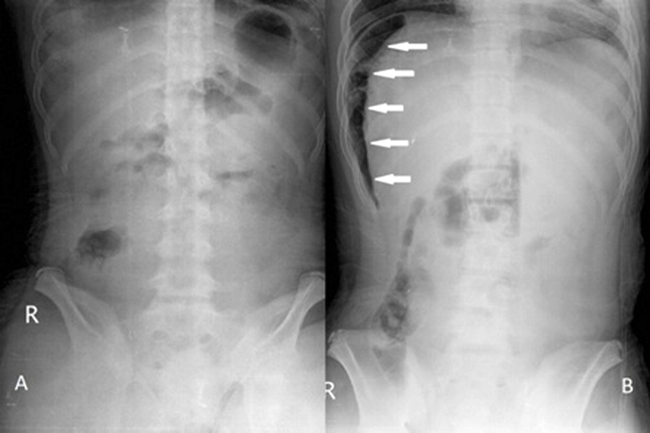
A 14-year-old boy presented to the emergency department with progressive abdominal pain for 1 week, which was epigastric, dull initially but then sharp in nature. Cold sweating, occasional vomiting, stressful life style were associated but no radiation pain was told. He was afebrile and denied drug consumption such as non-steroidal anti-inflammatory drug or Chinese herbs. At arrival, physical examination disclosed abdominal muscle guarding with board-like rigidity. Standing plain abdominal film showed no subphrenic free air (figure 1A). Perforated peptic ulcer with peritonitis was still highly suspected and further decubitus film revealed pneumoperitoneum (figure 1B, arrows). Timely laparotomy confirmed the diagnosis of perforated peptic ulcer with perforation over the anterior wall of pyloric ring. An erect chest or abdominal radiograph is the first choice in approaching acute abdomen and early detection of organ perforation is important. However, free intraperitoneal air is not always visible initially and the decubitus view may offer a more convenient, rapid, non-invasive, cheap and less radiation exposure investigating tool at the emergency department, comparing with the CT.1 In the era of high technology, the primary healthcare provider should be more confident of clinical judgment, even there is no air in the first radiograph.
(A) Plain film of abdomen showed no subphrenic free air. (B) The lateral decubitus view of abdomen revealed intraperitoneal free air (arrows).
References
Footnotes
-
Competing interests None.
-
Patient consent Obtained.