Article Text

Statistics from Altmetric.com
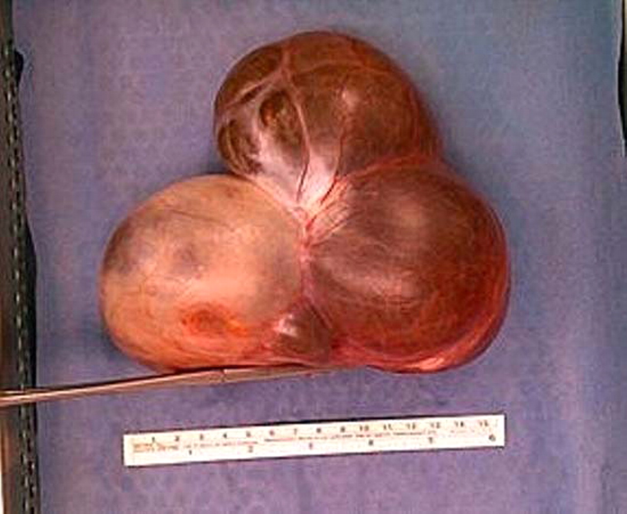
Description
An 18-year-old female presented to the emergency room complaining of atypical pain in the right hypocondrium. Clinical examination revealed the presence of an extensive mass in the right abdomen. Transvaginal ultrasonography (TVUS) detected the presence of a large anechoic, multilocular lesion (16×12×15 cm, with more than five locules), with septums <3 mm and colour score 1 (based on International Ovarian Tumor Analysis publication), which originated from the right ovary. The ultrasound findings were confirmed on CT scan (figure 1). At gynaecological laparotomy, a complex lesion consisting of multilocular elements and the falloppian tube was found. The latter was totally excised. Macroscopically, we observed a mass, which comprised the ovarian lesion and the tube, and which was characterised by lobularity (figures 2 and 3). Drainage revealed serous fluid. Histopathology showed characteristics of serous cystadenoma.

CT scan of a huge multiloculated adnexal mass.

Macroscopic view of the specimen of the right ovary which seems to be a large multiloculated mass.

Similar view of the right ovarian mass.
Ovarian cysts are found in transvaginal sonograms in nearly all premenopausal women and in up to 18% of postmenopausal women.1 Most of these cysts are functional in nature and benign. Ovarian cysts can be found in almost all prememopausal women and about 15% of postmenopausal women. The most common cysts are formed during the normal menstrual cycle and can be either follicular or luteal in origin. These cysts are benign and can range in size from 1 cm to 8 cm. Haemorrhage inside the cyst results in the so-called chocolate cyst. When an adnexal mass is suspected, TVUS is the imaging modality of choice. TVUS and serial measurements of the biomarker CA-125, have been included for the high-risk population.2 Ultrasound findings can help to distinguish between benign and malignant tumours. Other than ultrasound, CT or MRI is useful for larger masses and for examining the abdomen for metastases.3 Surgery is recommended for simple cysts larger than 5 cm and complex cysts of any size.
Acknowledgments
The authors thank the First Internal Medicine Department and the Ob/Gyn Department, Dr Karatapanis and Dr Sisamotos for this exellent work to be published.
Footnotes
-
Competing interests None.
-
Patient consent Obtained.