Article Text

Statistics from Altmetric.com
Description
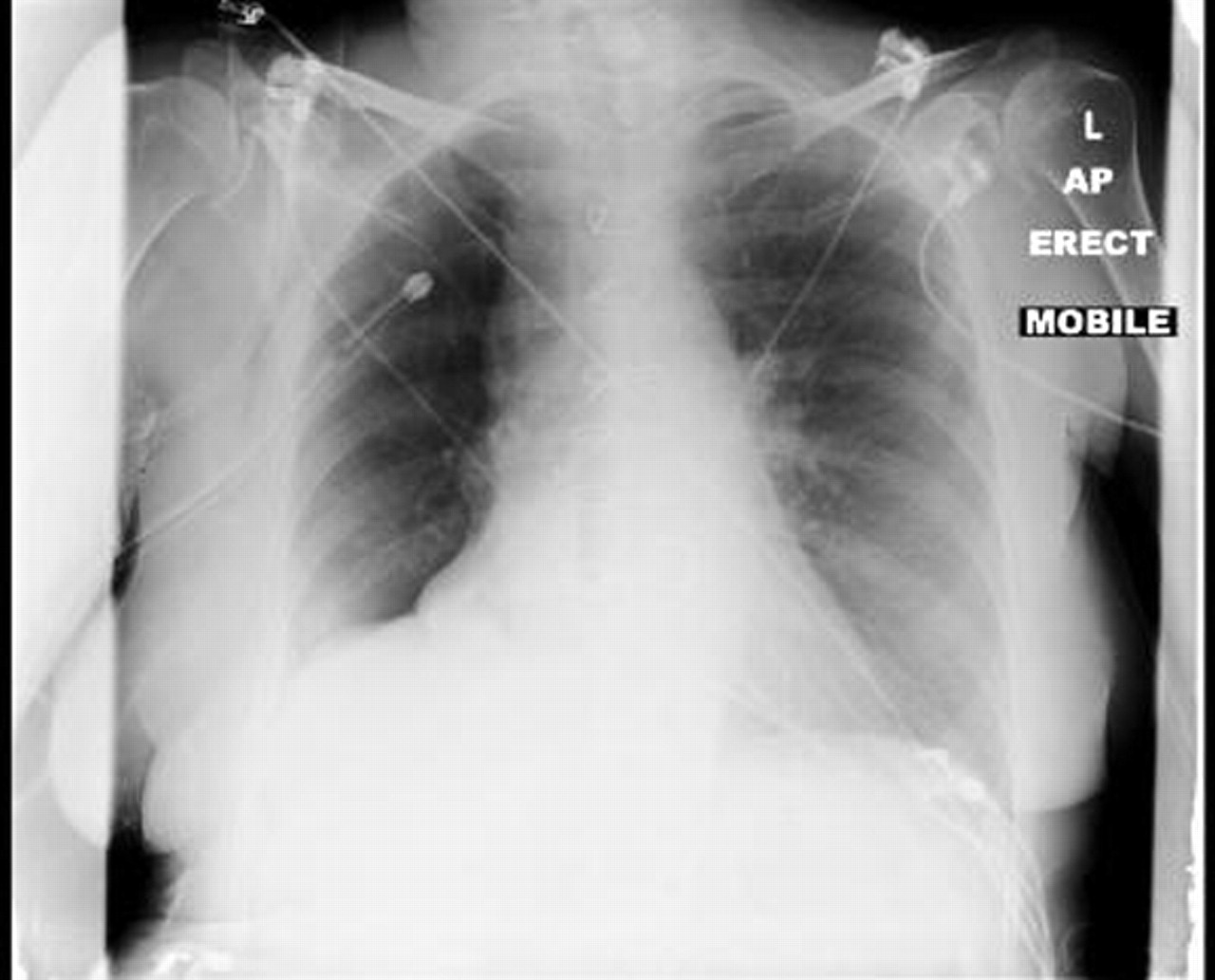
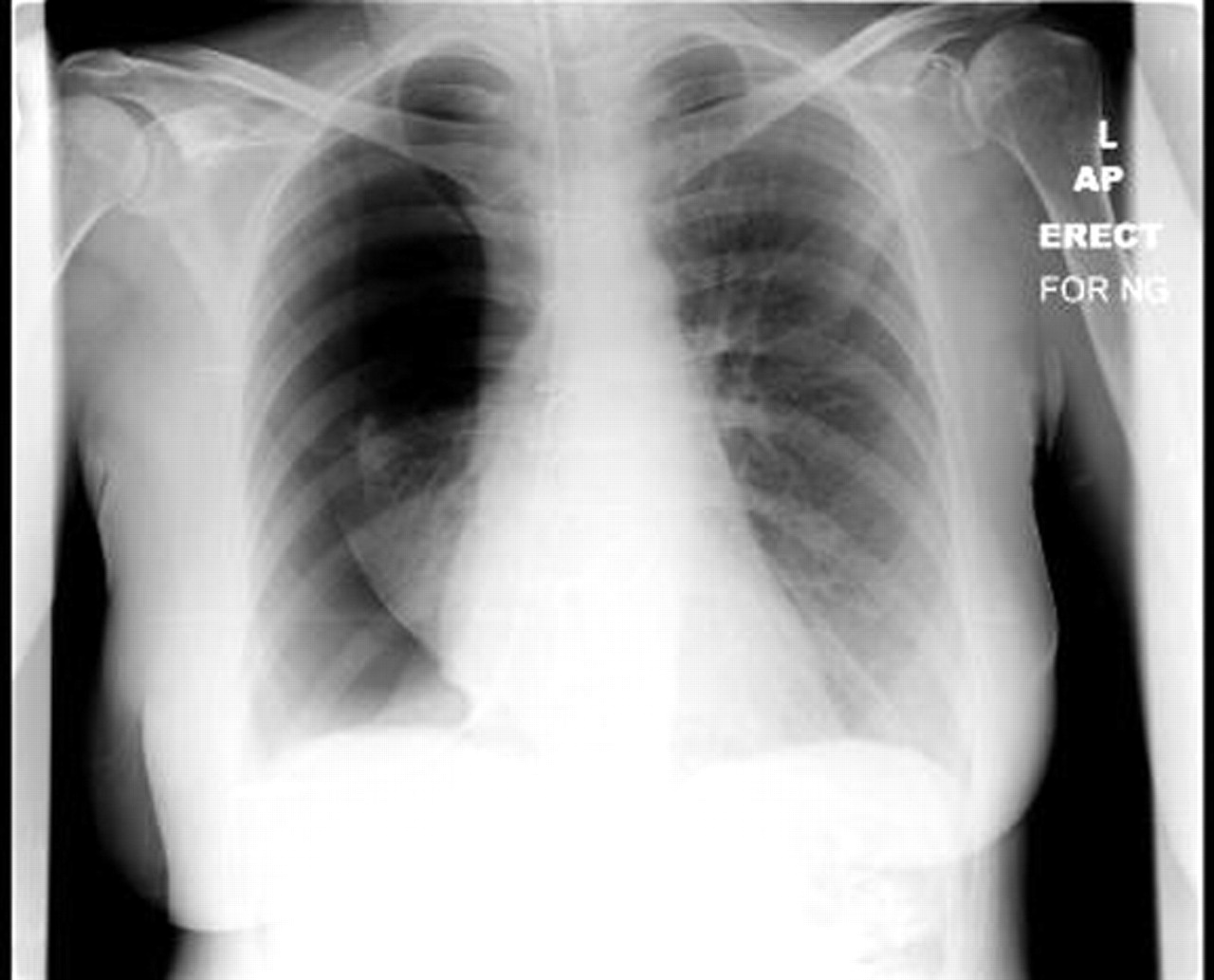
A 44-year-old woman with a background of a mitral valve replacement presented to the emergency department with aphasia. A CT scan of the brain demonstrated an infarction in the territory of the left middle cerebral artery. She was not suitable for thrombolysis and was admitted to the stroke ward where a nasogastric (NG) tube was passed to allow administration of her medication and nutrition. The check chest x-ray revealed that the NG tube had passed down the right main bronchus and through the lung causing a large pneumothorax (figure 1). A surgical chest drain was then inserted and a repeat radiograph showed re-inflation of the right lung (figure 2). This case demonstrates a serious complication of a common clinical procedure through inadvertent insertion of the NG tube into the airways. Studies have reported the incidence rate of accidental insertion into the trachea and smaller airways ranges from 0.3% to 15% and is more common in the older, mentally obtunded and patients who are intubated and sedated.1 2 Confirmation of gastric placement of the NG tube through clinical examination is unreliable and it is recommended that aspiration of gastric fluid and measuring a pH <5.5 should be obtained before commencing enteral feed.2 3 However, false positive results are possible as infected pleural or respiratory secretions can yield an acidic pH. A chest x-ray should be performed in patients who have a negative aspirate and the high risk groups described above.2 3

Nasogastric tube passed down the right main bronchus causing a large pneumothorax.
Re-inflation of the right lung after chest drain insertion.
Footnotes
-
Competing interests None.
-
Patient consent Obtained.