Article Text

Statistics from Altmetric.com
Description
An 83-year-old Jehovah's witness presented with a 4-month history of dysphagia and neck discomfort. She was known to have a thyroid adenoma, originally diagnosed back in the 1960s, but declined operative treatment at the time due to religious issues surrounding the need for a blood transfusion.
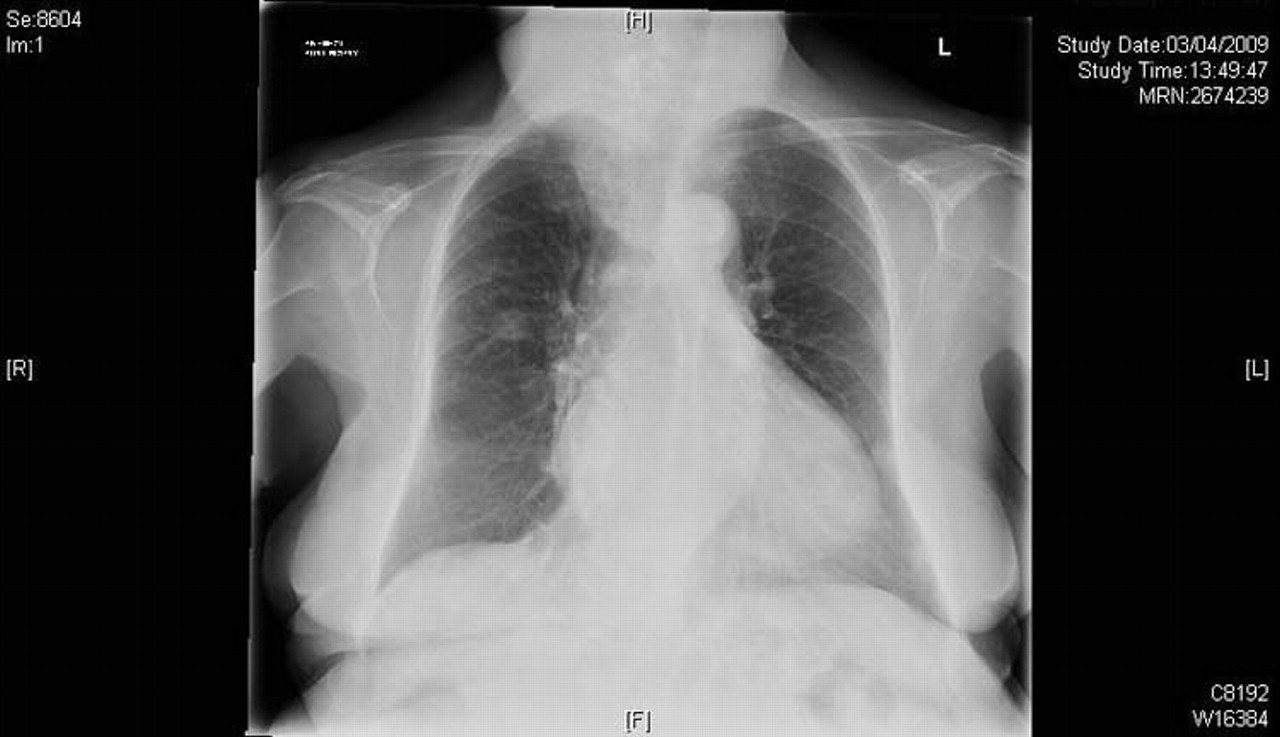
She was presently concerned about a progressive enlargement of the right side of her neck over the course of 6 months. Physical examination revealed a massive goitre. The upper and lower margins were not readily definable. Serum thyroid stimulating hormone and free thyroxine levels were normal. An urgent CT scan of her head and neck was ordered (figure 1). This demonstrated a significantly large thyroid mass with a necrotic centre and retrosternal extension. Her oesphagus was compromised and confirmed on oesophagogastroduodenoscopy. Chest x-ray confirmed tracheal deviation (figure 2).

CT of neck showing tracheal compromise.
Chest x-ray showing tracheal deviation.
Total thyroidectomy and neck dissection was performed and histology revealed fine, granular, calcified tissue, consistent with a thyroid adenoma. The patient's symptoms resolved following surgery.
The images are important because they demonstrate the significant size that thyroid masses can reach before they cause significant airway or oesophageal compromise especially if they are slow growing over many years.
Assessment of thyroid masses is threefold: clinically with regards to symptoms, on examination in terms of palpation and technically in terms of imaging and functional performance—that is, spirometry and endoscopy.
Footnotes
-
Competing interests None.
-
Patient consent Not obtained.