Article Text

Statistics from Altmetric.com
Description
A 38-year-old South-East Asian lady presented to breast clinic with a right peri-areolar breast lump. She denied any history of breast disease, breast surgery or significant family history. Examination revealed nodularity in the upper outer quadrants of both breasts and a more discrete right breast lump suggestive of a cyst. The impression was of fibrocystic disease, a common occurrence in this age group of women. Bilateral mammograms were performed (figure 1) which showed significant changes from those taken in 2009 following a presentation with right mastalgia (figure 2). Further questioning revealed that 12 months prior, she had collagen injected into her breasts to provide uplift and firming. This was brought into the UK from the Philippines and injected by a friend, who was inexperienced in this field. Such practice is common throughout Asia, particularly the Philippines. Collagen, in the form of recombinant human or bovine collagen, is one of the most commonly used fillers for soft tissue augmentation.1 Although histological evidence of collagen in soft tissue disappears 6 months postinjection,2 no literature evidence exists documenting long term effects of its use in the breast, especially in the case of injection in inexperienced hands. In light of this, we intend to follow-up and re-image the patient in 1 year. With awareness of this practice, it is important that we are able to recognise the appearance of collagen on mammography and be vigilant of how such appearances may obscure underlying abnormalities, possibly reducing the sensitivity and specificity of breast screening.
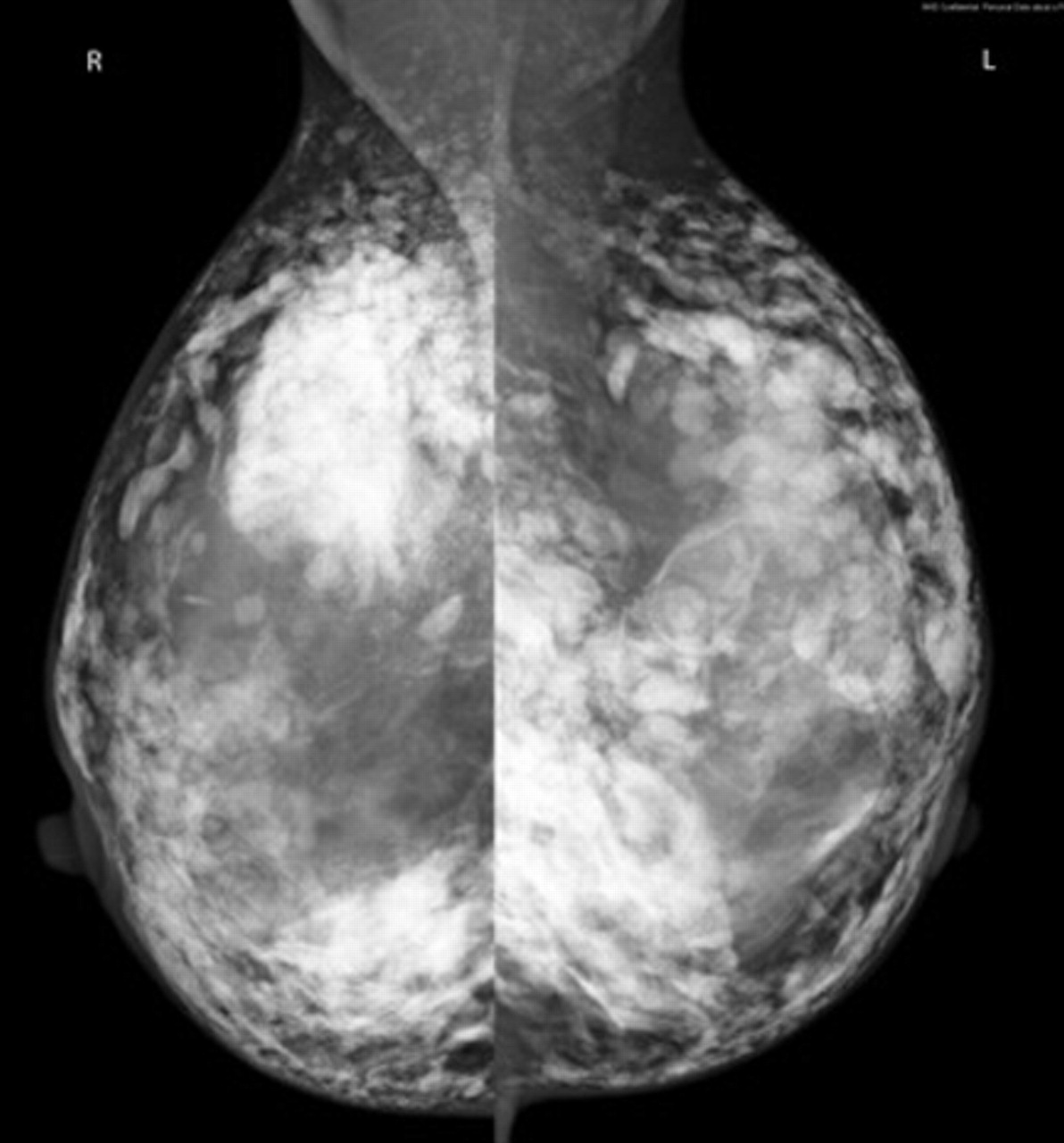
Multifocal increased densities with a rounded nodular appearance consistent with injection into both breasts.

Moderately dense glandular breast tissue with no focal abnormality.
Acknowledgments
Many thanks to Mrs Susan Jones for her expert help and guidance.
Footnotes
-
Competing interests None.
-
Patient consent Obtained.