Article Text

Statistics from Altmetric.com
Description
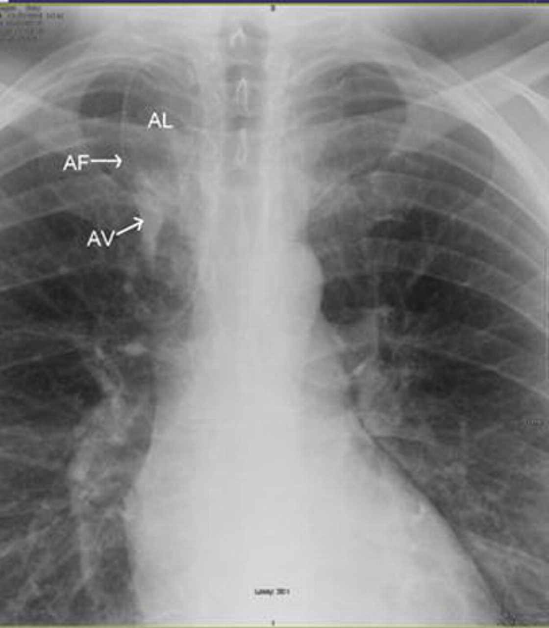
A 38-year-old male admitted to the hospital with chest pain for few days. He does not have any significant medical and surgical problems. Physical examination, lab work, imaging studies and exercise cardiolite stress were normal. Eventually, we concluded that this chest pain was most likely secondary to musculoskeletal origin. We found azygos lob incidentally on chest x-ray and computed axial tomography of chest. These images have typical appearance of azygos lobe, fissure and vein. The azygos lobe (azygos lobe in figures 1 and 2) is a uncommon anomaly that is found in 1% of anatomic specimens,1 on about 0.4% of chest radiographs2 and 1.2% of high resolution CT.3 The azygos lobe is a developmental anomaly but not a true accessory lobe. On chest radiographs, azygos fissure (azygos fissure in figure 1) is visible as a fine convex line and its upper portion has triangular shape due to extrapleural areolar tissue and lower portion has tear-shaped shadow due to azygos vein (azygos vein in figure 1). Anatomy of azygos lobe is clinically important during thoracic surgical procedures. Cases of spontaneous pneumothorax have reported in patients with azygos lobe.

Chest x-ray image.

Computed axial tomography image. AL, azygos lobe; AF, azygos fissure; AV, azygos vein.
Acknowledgments
The authors would like to thank Dr Martin Joshua G MD for his contribution.
Footnotes
-
Competing interests None.
-
Patient consent Obtained.