Article Text

Statistics from Altmetric.com
Description
A man presented with a 1-year history of a progressively worsening penile skin lesion. Over the preceding 2 months, he reported passage of urine from two distinct exit points on the distal penis. There were associated low grade penile pain, dysuria, spraying of urine on micturition, daytime urinary frequency, nocturia of three, the sensation of incomplete bladder emptying and contact bleeding at the distal penis. On presentation, there was a 6 cm circumferential fungating lesion on the distal penis. There was evidence of superficial spreading. The lesion extended 8 cm proximally. There was evidence of ulceration and contact bleeding. It was not possible to identify the external urethral meatus (figures 1 and 2). There was no associated inguinal lymphadenopathy. Biopsy showed invasive, well differentiated squamous cell carcinoma. An inguinal ultrasound was negative for lymphadenopathy. A staging CT scan of the thorax, abdomen and pelvis showed no evidence of metastatic disease. A total penectomy and formation of a perineal urethrostomy was performed (figures 3–5). The pathological specimen showed an invasive, well differentiated squamous cell carcinoma. The tumour measured 9 cm in length and 6 cm in diameter. The carcinoma invaded into the subepithelial connective tissue and the underlying corpus cavernosa. There was no evidence of vascular or urethral invasion. The proximal resection margins were free of tumour. The specimen was classified as pT2 N0 M0. The patient made an uneventful recovery. Routine follow-up is scheduled at 6 months intervals for the first 2 years, and yearly thereafter to 2 years. It shall consist of a thorough physical examination.

Preoperative anterior view of the fungating penile lesion.
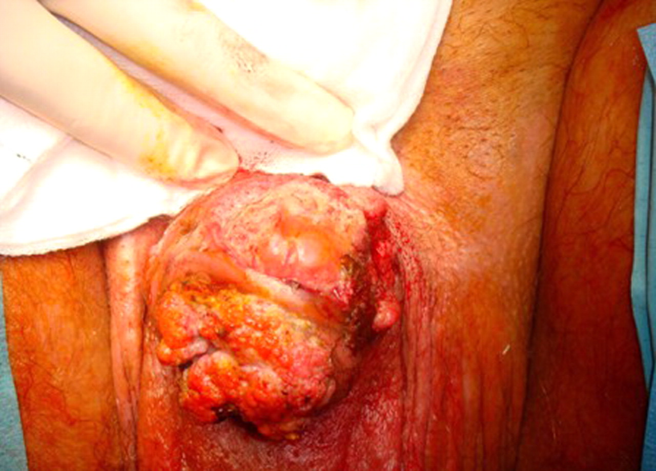
Preoperative anterosuperior view of the fungating penile lesion.

Anterosuperior view of the resected penile lesion. The specimen measured 16 cm in length with a diameter of 6.5 cm and 19 cm circumference.

Lateral view of the total penectomy specimen.

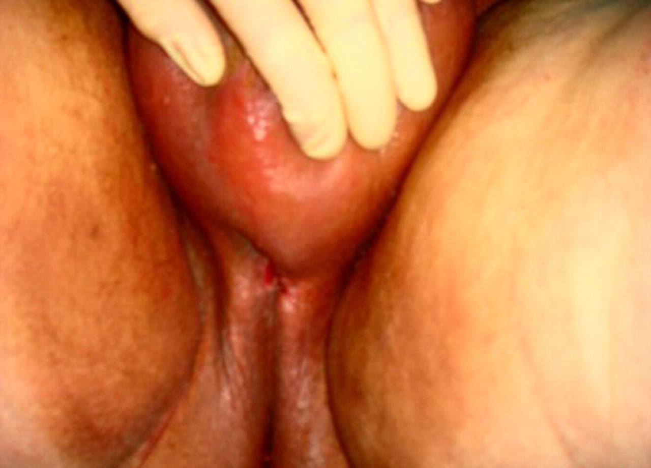
Anterior view of the perineal urethrostomy at day 7 post-total penectomy.
Footnotes
-
Competing interests None.
-
Patient consent Obtained.