Article Text

Statistics from Altmetric.com
Description
Mature cystic teratoma is the most common benign ovarian neoplasm occurring in the reproductive age group.1 Sonography is invariably the primary modality for diagnosis. The great diversity in components of teratomas results in varied sonographic appearances. Certain features like tip-of-iceberg sign, fat-fluid level, dermoid mesh and rokitansky protuberance (dermoid plug) are specific to this entity and considered diagnostic.2 A sonographic appearance of multiple free-floating spherical balls in a cyst as seen in the presented case is rare2 and may present a diagnostic difficulty to the unwary.
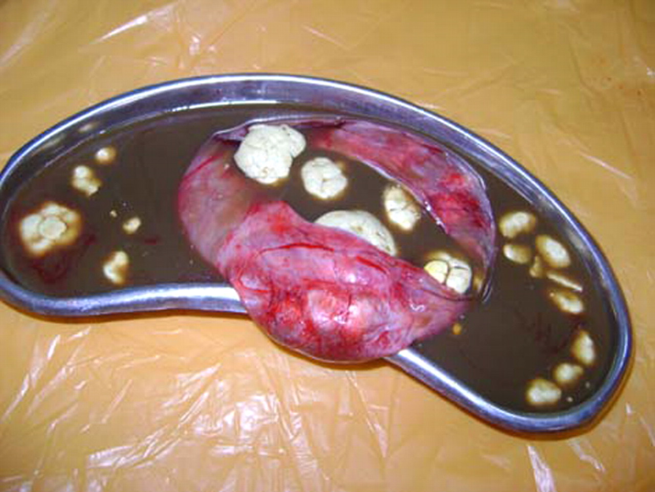
Transabdominal sonography in a 23-year-old patient complaining of lower abdominal discomfort revealed a 12×8 cms cystic mass in the lower abdomen. The cyst had a striking ultrasound appearance with multiple distinct, round 1–2 cm sized echogenic globules floating in it (figure 1). CT scan demonstrated a thin-walled cyst with hypodense floating balls within (figure 2). At laprotomy, a unilocular right ovarian cyst containing serous fluid and numerous small cheese-like sebum balls was found (figure 3). Histopathology confirmed the diagnosis of mature cystic teratoma; spherules contained desquamated keratin, fibrin, hemosiderin and sebaceous debris with skin squams and fine hair shafts and only a small amount of fat component.
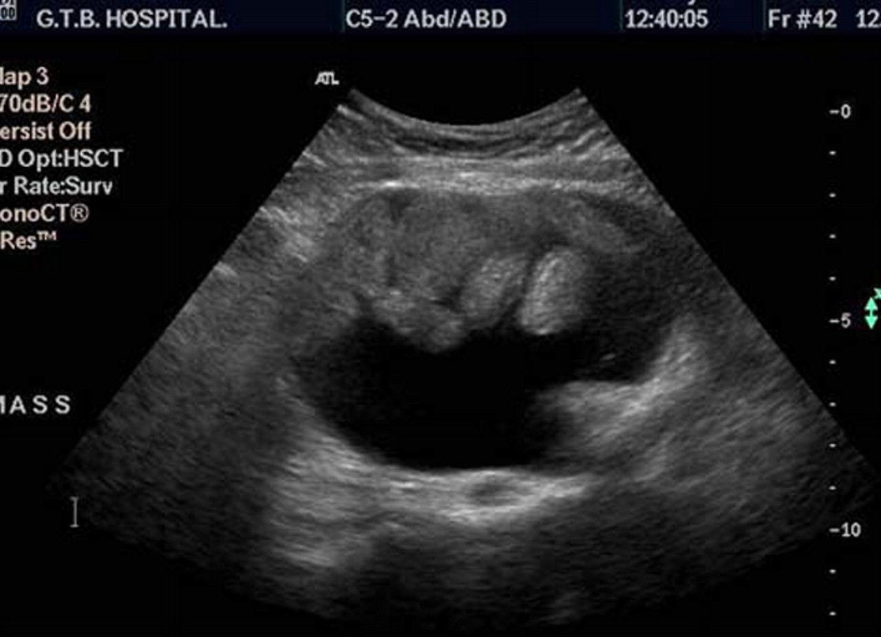
Sonogram showing multiple echogenic floating spherules in a cyst.

Axial CT image shows a thin-walled cyst with hypodense floating balls within.
Gross specimen of the ovarian cyst showing multiple cheese-like sebum balls in serous fluid.
The mechanism of formation of these globules is unclear till date. It is postulated that globules are formed by aggregation of sebaceous material around a nidus while moving in the cyst resulting in concentric layers of sebaceous material. The lower specific gravity than cyst contents accounts for their mobility.3
Sonography is diagnostic and further workup is unlikely to show any additional features. To the best of our knowledge, this appearance has not been reported in other tumours and is pathognomic for mature cystic teratoma.
Footnotes
-
Competing interests None.
-
Patient consent Obtained.