Article Text

Summary
The authors describe the case of an athlete from the Brazilian national men’s basketball team (sub-16) who reported pain in the right iliac region at the end of the season. Clinical and imaging exams revealed an iliac bone stress fracture. A conservative treatment of removing the load from the fracture in combination with physical therapy was chosen. The athlete improved satisfactorily and returned to the sport at the same level as prior to the injury after 14 weeks of treatment.
Statistics from Altmetric.com
Background
Stress fractures are common among athletes and represent about 10% of overuse injuries in sports. The lower extremities are affected more frequently. Aetiological factors can be intrinsic (age, gender, skeletal alignment, hormonal factors, nutritional factors and bone density) and/or extrinsic (training, footwear and the sport).1
Approximately 69% of stress fractures occur in runners, with the most frequently injured areas being the tibia (34% of cases), distal fibula (24%), metatarsals (18%), femoral neck and shaft (14%) and pelvis (6%).2
Fractures that are considered low-risk (fractures of the first to fourth metatarsals, medial cortical fractures of the tibia, femoral diaphyseal fractures and medial cortical fractures of the femoral neck) have a favourable prognosis. These fractures are located in areas of bone compression, evolve with a low frequency of complications and improve with changes in sporting activities.
The areas that are considered high-risk (olecranon, a lateral cortical fracture of the femur, patella, anterior tibial diaphysis, medial malleolus, navicular, medial sesamoid and fifth metatarsal bone) have unfavourable outcomes and progress with a high frequency of complications, such as relapses, pseudoarthroses and complete fractures. Moreover, fractures in these areas frequently require surgical treatment.
Iliac stress fractures are rare and associated with bone insufficiency. Fractures in this area correspond to 4% of pelvic stress fractures, with two cases described in the literature to date.3 4
Case presentation
We describe the case of a 16-year-old male athlete from the Brazilian national basketball team (sub-16). During the South American championship, the athlete reported pain in the right iliac region that had no precipitating trauma but progressively worsened during sports practice. The pain primarily occurred when performing movements involving jumping and running. He reported improvement with rest but did not interrupt his activities. With 2 months remaining until the end of the championship, he played but did not train. He had been playing and training for an average of 3 h per day and six times per week for 6 years. He did not report any previous injuries. At the end of the season, the athlete was subjected to a medical evaluation. He reported a worsening of the pain, which, at that point, was occurring daily and continuously, even outside of sports practice. At the physical examination, he presented with claudication. He reported pain when supporting his lower right leg with only his right foot, with no antalgic posture. In addition, he presented with diffuse pain upon palpation of the right iliac region and upon right hip flexion against resistance. A neurovascular examination revealed no abnormalities (figure 1).
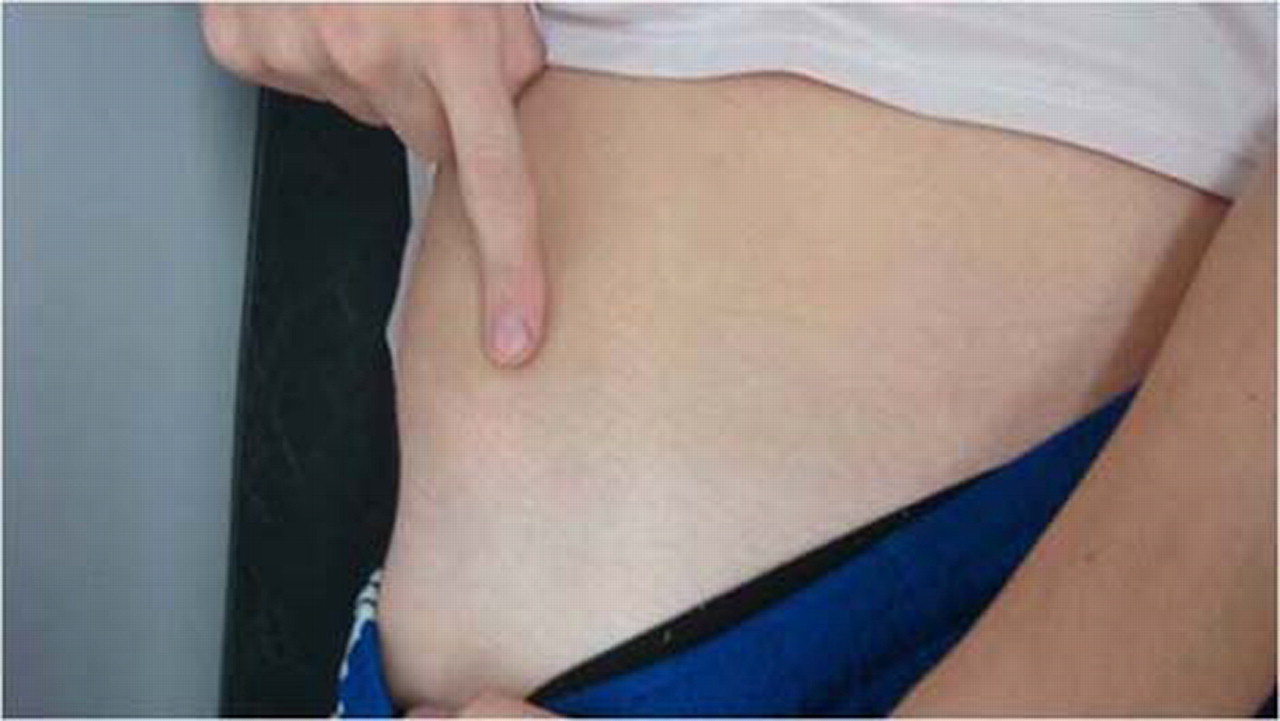
Localisation of the pain reported by the patient.
Radiography revealed no abnormalities, whereas nuclear MRI revealed irregularity and oedema in the bone marrow of the anterior superior iliac spine and apophyseal nucleus. An examination revealed a fracture line on the wing of the iliac bone, which could be seen from the coronal plane and had no fragmentation or significant misalignment (figure 2).

(A,B) Magnetic resonance image of a coronal section showing the iliac bone fracture line.
Due to the symptoms, a conservative treatment was chosen. The athlete was completely removed from his physical activities and advised to use crutches to avoid loading the right leg. In addition, analgesic physical therapy was initiated (figures 3 and 4).

(A,B) Magnetic resonance image from January 2009 showing fracture progression.

(A,B) Magnetic resonance from February 2009 showing fracture consolidation.
Differential diagnosis
▶ Muscular injury
▶ Tendinopathy
▶ Anterior superior iliac spine avulsion fractures
▶ Iliac bone apophysitis
▶ Iliac bone stress fracture
▶ Labral hip injury
▶ Femoroacetabular impact.
Outcome and follow-up
The athlete rested from all activities for 40 days. In January 2009, he began stretching and hydrotherapy. After 60 days, the pain improved, and he began muscular strength training, after which he progressively returned to lower-limb strength training and abdominal and aerobic training. After 14 weeks, he returned to the sport at the same activity level as prior to the injury.
Iliac bone stress fractures are uncommon injuries and are part of the differential diagnosis for skeletally immature patients. In the case described here, the aetiology was extrinsic. The athlete played at a very competitive level and was subjected to repetitive microtraumas, which produced stress that was excessive for normal bone and altered bone formation and reabsorption.5,–,7
Initial propaedeutic imaging is performed via simple radiography. However, radiography did not reveal a significant callus fracture in the region.8 MRI is the tool of choice because of its usefulness in identifying bone oedema in fracture areas.7 8
In this case, conservative treatment produced favourable results, enabling a return to the sport at the same level as prior to the injury.3 9
Learning points
▶ Iliac bone stress fractures should be considered during the differential diagnosis of hip injuries in athletes.
▶ In immature skeletons, an increase in the intensity and frequency of training can lead to injuries due to overuse.
▶ Non-surgical treatment should be the treatment of choice because it presents favourable clinical results.
Footnotes
-
Competing interests None.
-
Patient consent Obtained.
Linked Articles
- CORRECTION