Article Text

Statistics from Altmetric.com
Description
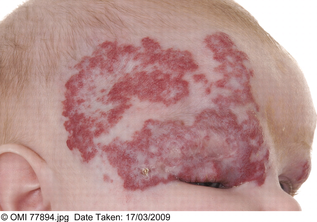
A 5-month-old female infant with a facial haemangioma (fig 1) presented with left focal seizures. Magnetic resonance (MR) imaging of the brain revealed ischaemic changes to both anterior and posterior territory circulations of the right hemisphere (fig 2). MR angiography showed gross abnormality of the cerebral vasculature, characterised by large vessel stenosis on the right with associated prominent perforators and also a congenital abnormal left carotid system (fig 3). These radiological features, in combination with the cutaneous lesion, was suggestive of PHACE syndrome.1,4

Right facial large segmental haemangioma.

Right anterior and posterior circulation infarcts.

Very thin right middle cerebral artery (MCA) and internal carotid artery (ICA) abnormally arising directly from aortic arch.
This infant had initially presented at 1 month of age with an enlarging right segmental facial haemangioma which was managed with oral prednisolone and surveillance by secondary care providers.
At the 6 week baby check the general practitioner had commented on a circumscribed skin lesion on the sternum (fig 4) which in itself had not prompted further investigation.

Sternal defect.
In 1996 Frieden first described PHACE syndrome—a heterogeneous neurocutaneous disorder that includes posterior fossa malformations (P), facial haemangiomas (H), arterial anomalies (A), cardiac anomalies (C), and eye abnormalities (E).1 Recent authors include sternal defects and refer to the syndrome as PHACE(S).3 Our infant was found to have the facial haemangioma (H), (cerebral) arterial anomalies (A), and sternal defect (S).
This presentation is consistent with the observation that few infants manifest with the entire constellation of anomalies. The cerebrovascular anomalies are of concern because of the risk of early infantile stroke. This case reiterates assertions made by other authors that the presence of a characteristic segmental facial haemangioma necessitates evaluation for the extracutaneous features of PHACE(S) syndrome.2
Footnotes
Competing interests: None.
Patient consent: Patient/guardian consent was obtained for publication.