Article Text

Statistics from Altmetric.com
Description
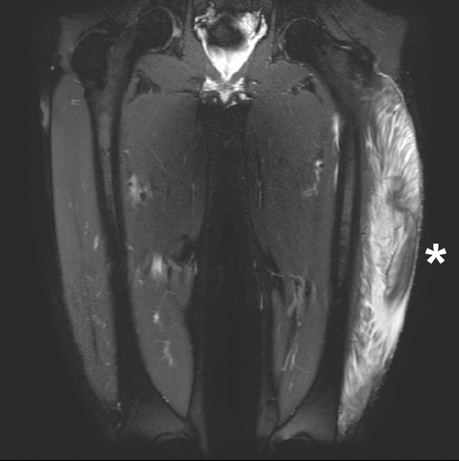
A previously healthy, 45-year-old man presented with a 2 day history of increasing pain in the left thigh, which was swollen and tender, without any history of trauma. Blood pressure, peripheral pulses and laboratory tests were normal. Compartmental pressure in the anterior left thigh was 50 mm Hg (normally <30 mm Hg), indicating an acute compartment syndrome (ACS).1 Magnetic resonance imaging (MRI) examination, obtained in order to exclude a rapidly growing tumour, showed a non-enhancing mass in the anterior compartment of the thigh (figs 1 and 2), representing an acute intramuscular haematoma and widespread muscular oedema, suggesting ACS. An immediate fasciotomy was performed whereby 300 ml of coagulated blood was evacuated from the vastus lateralis. The fascia lata was then easily sutured. The patient was discharged and quickly recovered.

Coronal STIR (short tau inversion recovery) magnetic resonance image (MRI) of the thighs, showing a large intramuscular haematoma (asterisk) as well as widespread muscle oedema in the ventral compartment of the left thigh.

Axial T2 weighted MRI of the thighs, showing a large intramuscular haematoma (asterisk) as well as widespread muscle oedema in the ventral compartment of the left thigh.
Risk factors for ACS include: systemic hypotension, history of external compression or trauma (with/without fracture), coagulopathy, and vascular injury.1 Retrospectively, our patient recollected having suffered a minor blunt trauma to the thigh, playing with his children, 2 days earlier.
Morbidity from ACS is high,2 and its outcome depends upon the balance between local blood pressure and compartment pressure as well as on timely decompression.2 Most patients benefit from early fasciotomy, where the risks and sequelae are much less serious than those of untreated ACS2
Early diagnosis supported by measurement of compartmental pressure is mandatory for a successful therapeutic outcome of ACS, irrespective of location. Imaging can be useful for differential diagnosis.3 Immediate fasciotomy is indicated to prevent neurovascular compromise and tissue necrosis with or without systemic consequences and functional impairments.
Footnotes
Competing interests: None.
Patient consent: Patient/guardian consent was obtained for publication.