Article Text

Statistics from Altmetric.com
Description
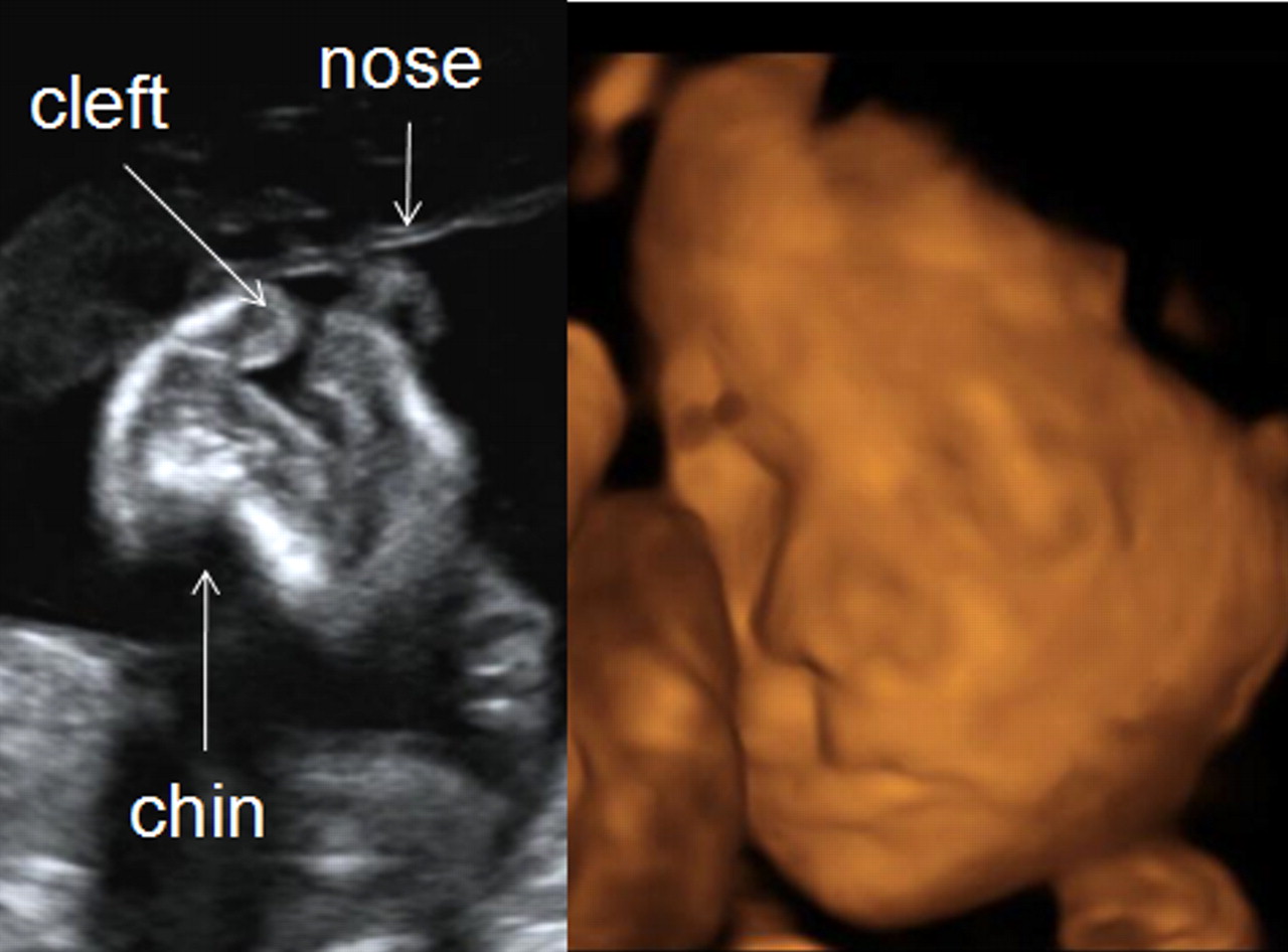
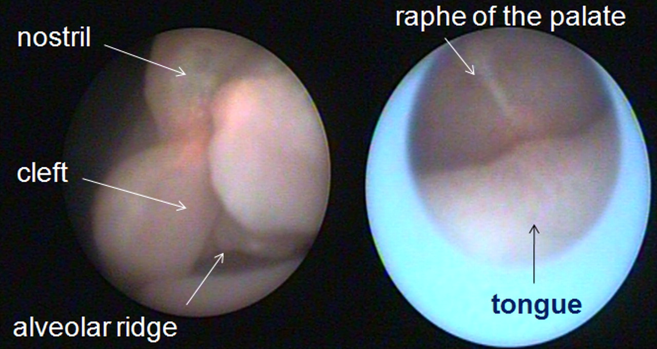
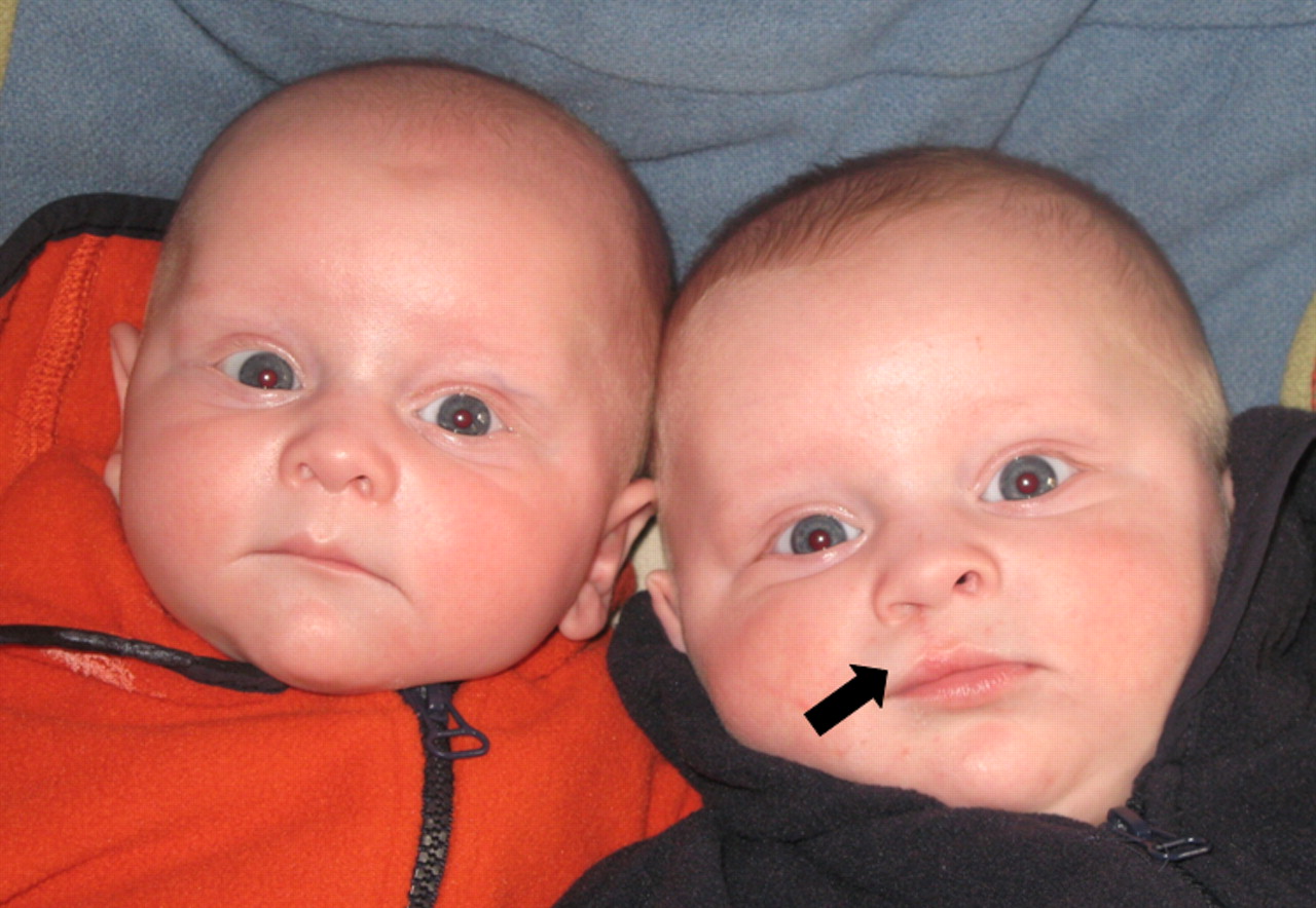
A 32-year-old woman, 22 weeks pregnant of monochorionic diamniotic twins, underwent fetoscopic laser coagulation of placental vascular anastomoses for a twin-to-twin transfusion syndrome (TTTS) Quintero stage III. Preoperative two- and three-dimensional ultrasound revealed the presence of an isolated unilateral cleft lip in the recipient twin (figure 1). At the time of surgery, fetoscopic inspection of the upper lip confirmed the unilateral cleft and the intact raphe of the palate (figure 2, video 1). The postoperative course was favourable and the patient delivered at 36 weeks of gestation. The cleft lip was surgically repaired soon after birth. Now aged one, both twins do fine and the remnants of the cleft can hardly be seen (figure 3).

Two dimensional (left) and three dimensional (right) ultrasound image of fetal cleft lip.

Fetoscopic image of cleft lip (left) and intact palate (right).
Fetoscopic investigation of a cleft lip.

Postnatal image after surgical reconstruction of the lip. The scar in the affected twin (arrow) can only hardly be visualised.
Anomalies of the midline structures occur more often in monochorionic twins than in singletons, probably due to the zygotic clefting. In 6% of cases this malformation is discordant.1 As shown here, the prognosis of an isolated cleft lip is generally good and should therefore not influence the treatment of TTTS.2 We used the fetoscopic laser coagulation as an opportunity to visualise the recipient fetus directly and to confirm the morphological anomaly. Historically, embryoscopy has also been used by others as a complementary tool to assess birth defects early in pregnancy.3 However, this had high complication rates and was abandoned for the assessment of clefts, especially since (3D) ultrasound allows adequate description of fetal lip and palate. Animal experiments are now ongoing to assess the role of fetoscopy in the prenatal therapy of facial clefts.4
Footnotes
-
Competing interests None.
-
Patient consent Obtained.