Article Text

Statistics from Altmetric.com
Description
Figure 1 shows a fungating but no-cancerous breast tumour. Rapid growth of the non-malignant stromal tumour described as a phyllodes tumour involved most of the breast and caused breast ‘rupture’ from pressure necrosis of the overlying skin.
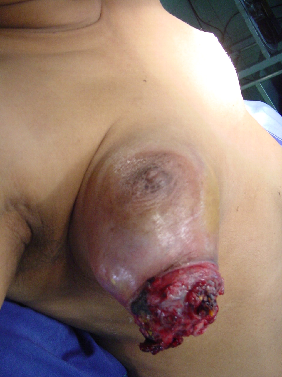
The right breast showing a large fleshy tumour protruding from the lower outer quadrant with most of the remaining breast also containing tumour. The surrounding skin although bluish is not oedematous.
The 48-year-old woman had been institutionalised and poorly cared for because of schizophrenia. A small breast lump had been present over many years and had started to enlarge in the last 3 months. The late presentation was due to social withdrawal, paranoia and refusal to seek treatment.
She was single and nulliparous but without a family history of breast cancer. Fungating tumours of the breast are commonly attributed to invasive duct carcinoma. An everted ulcer edge and oedema of the surrounding skin are characteristic. Nevertheless, images presented show a tumour without skin oedema, fixity or attachment suggestive of malignant infiltration. There was no axillary lymphadenopathy or distant metastases. Core biopsy revealed leaf-like architecture with marked stromal overgrowth and hypercellularity suggestive of a borderline phyllodes tumour (figure 2). Clonal studies have confirmed that fibroadenomata could progress to phyllodes tumours.1 These tumours tend to be locally invasive, but only rarely metastasise to distant sites. Wide local excision alone is recommended to prevent local recurrence.2 3

Histology with haematoxylin eosin stains revealed leaf-like architecture, long clefts with marked stromal overgrowth and hypercellularity characteristic of a phyllodes tumour.
Aggressive pharmacotherapy and counselling enabled optimum treatment in this instance. Considering the large size of the tumour she underwent simple mastectomy with clear margins and remains disease-free 4 years later. She now lives with her sister and complies with regular follow-up medical care.
Learning point
Rarely, long standing fibroadenomata transform in to locally aggressive stromal tumours known as phyllodes tumours and proceed to ulcerate without skin infiltration—that is, nodules, peau d'orange.
Poorly managed psychiatric disorders may lead to late presentation and delayed treatment.
Enucleation is insufficient and wide local excision is recommended to prevent local recurrence.
Footnotes
-
Competing interests None.
-
Patient consent Obtained.