Article Text

Statistics from Altmetric.com
A 78-year-old woman with long-standing bilateral knee osteoarthritis underwent a right total knee arthroplasty(TKA). She had previous history of hypertension and breast cancer. There were no complications during the operation and the pedal pulse was present after TKA. On the fifth day after TKA, the examination revealed pain in the right calf that increased when she walked, swelling and oedema in the popliteal fossa, local haematoma and a pulsatile mass. All extremity pulses were palpable.
The duplex showed a 3 cm hypoechoic mass in the popliteal fossa with Doppler flow consistent with a popliteal artery pseudoaneurysm and no signs of arteriovenous fistula. A limb arteriogram (figures 1–4) confirmed a pseudoaneurysm arising from the third portion of the popliteal artery just below the tibial prosthesis.
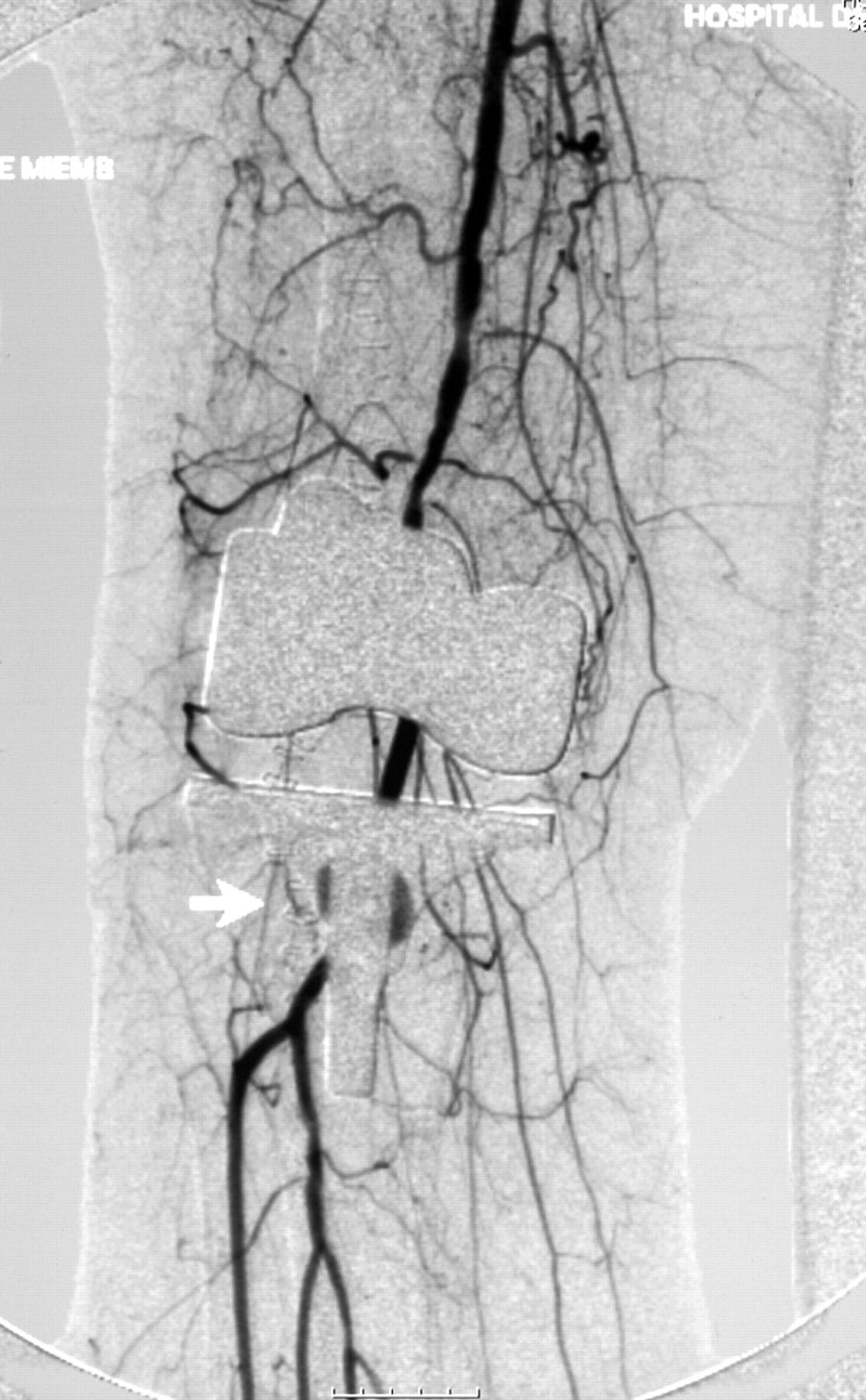
Digital subtraction antero-posterior arteriogram showing a pseudoaneurysm arising from the third portion of the popliteal artery just below the tibial prosthesis.

Frontal view.
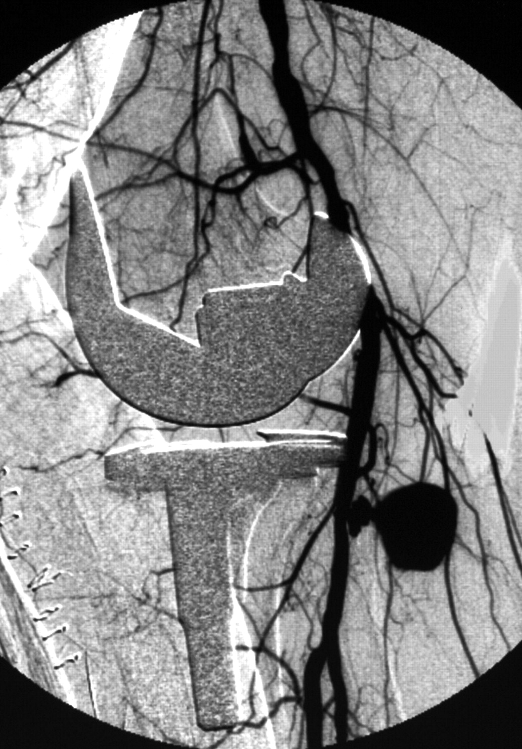
Digital subtraction lateral arteriogram showing a pseudoaneurysm arising from the posterior wall of popliteal artery at the joint level of the knee prosthesis.

Pseudoaneurysm with the run-off tibial arteries.
The pseudoaneurysm was urgently excised, open-surgically repaired with direct suture of the bleeding point and the haematoma drained. Surprisingly, a well-shaped circular hole was identified only on the posterior wall of the artery. Distal pulses were present at the end of the vascular surgery.
The patient had no further complications. The postoperative duplex showed no evidence of residual pseudoaneurysm and after 48 h the patient was walking without difficulty.
This case illustrates an arterial complication after TKA, which is infrequent (incidence ranges from 0.03–0.5%).1
It is very important to perform a prompt diagnosis and treatment to avoid serious complications like limb loss.2
Treatment includes conservative management, open-surgical repair, and embolisation or endovascular stenting. The latter is the less desirable treatment because it is associated with a high rate of early occlusion.3
Footnotes
Competing interests: None.
Patient consent: Patient/guardian consent was obtained for publication.