Article Text

Statistics from Altmetric.com
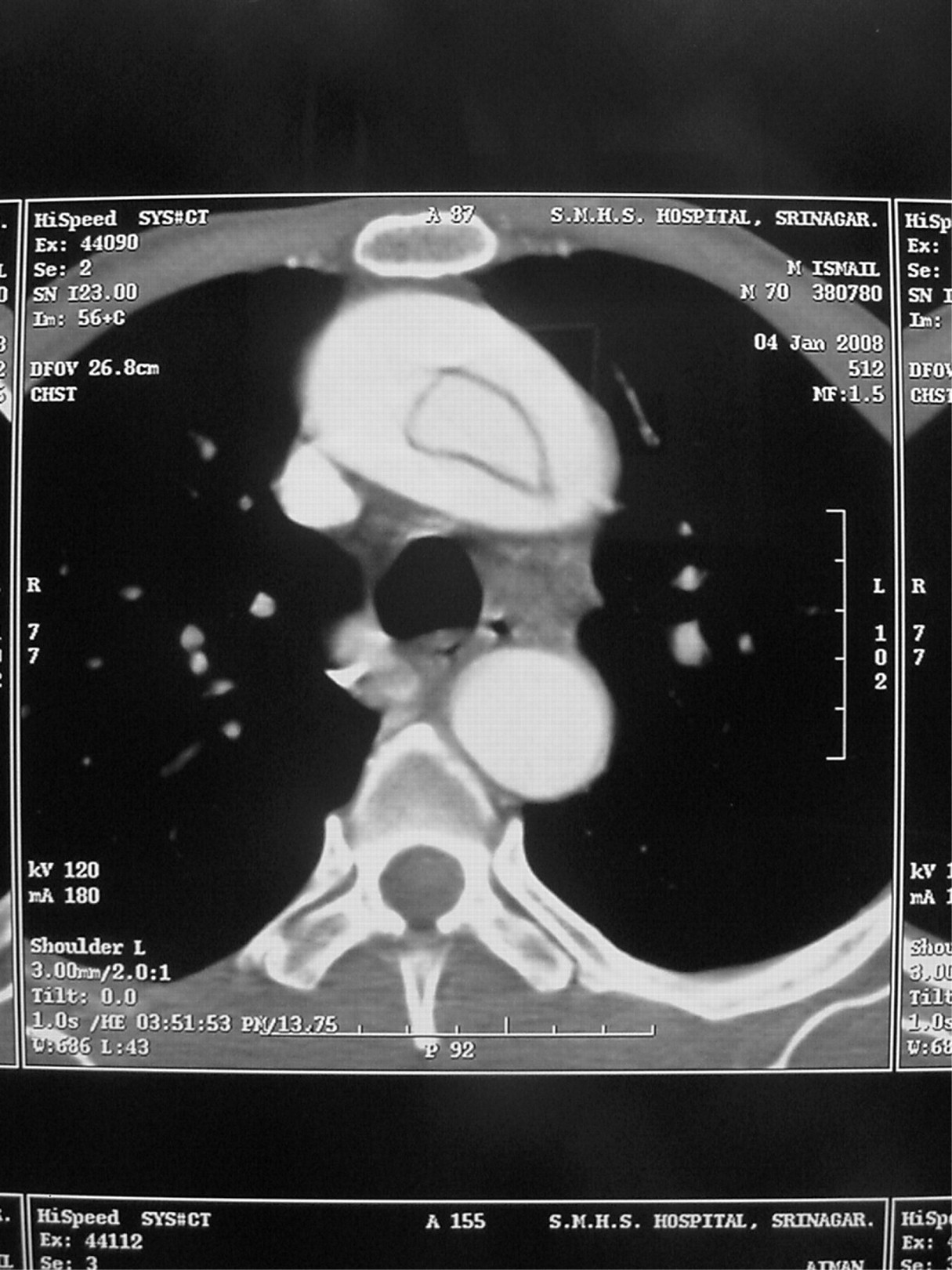
An adolescent otherwise healthy male presented with acute onset chest pain. Physical examination was remarkable for early diastolic murmur of aortic regurgitation. Transthoracic and subsequently transoesophageal echocardiography revealed aortic dissection extending to the arch. CT angiography revealed a circumferential type of aortic dissection extending from the aortic root to the arch (type A) with extension of the dissection flap into the right brachiocephalic artery (fig 1).
DISCUSSION
The typical aortic dissection appears on contrast enhanced CT as an intimal flap that separates the false from the true lumen. Features indicative of a true lumen are outer wall calcification and eccentric flap calcification; beak sign, larger cross sectional area and slow enhancement are indicators of a false lumen.1–4 In some instances, an atypical configuration of intimal flap is encountered. These include a calcified false lumen in chronic cases, an aorta with three or multiple channels, an extremely narrow true lumen, or rarely, as in the present case, a circumferential intimal flap.1 3 The latter may be further complicated by intimointimal intussusception, although this was not evident in our case.5
Acknowledgments
This article has been adapted from Choh N A, Choh S A, Naikoo B, Bhat S A, Jehangir M. Circumferential aortic dissection Archives of Disease in Childhood 2008;93:581
Footnotes
Competing interests: None.