Article Text

Statistics from Altmetric.com
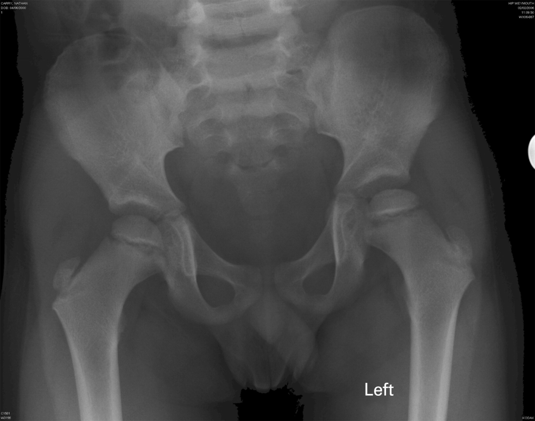
A 6-year-old boy was referred to the regional paediatric rheumatology service with a 1-year history of limp.
The patient was initially thought to have an irritable hip and x rays including the frogs legs view requested by the GP were reported as normal. In view of this no action was taken. However, his symptoms persisted and he was referred to a paediatric rheumatologist. On examination he had severe restriction of internal rotation and a clinical diagnosis of Perthes disease was suspected despite the normal x ray. An MRI showed partial collapse of the left femoral head with areas of necrosis, confirming the diagnosis of Perthes disease.
x Ray appearances may be normal in the early stages of Perthes disease, hence continuing symptoms warrant either a repeat x ray after 3–4 weeks or an MRI scan.


Acknowledgments
This article has been adapted from Meadows C, Monsell F, Ramanan A V. Normal x rays in Perthes disease Archives of Disease in Childhood 2008;93:211