Article Text

Statistics from Altmetric.com
A 25-year-old woman presented in altered sensorium that developed after an episode of complex partial seizure 6 days ago. She had a history of low-grade fever, anorexia and weight loss for 6 months. She also had productive cough for 4 weeks. She was febrile and breathing fast. Her neck was supple and no meningeal signs were present. Deep tendon reflexes were hyperactive and fundus examination was normal. She had generalised lymphadenopathy.
Serum electrolytes were normal. Lumbar puncture was performed and cerebrospinal fluid (CSF) opening pressure was normal. CSF examination showed increased proteins, normal glucose and lymphocytic pleocytosis. On the basis of a history of high-risk sexual exposure, HIV ELISA was performed and was reactive. Her CD4 count was 90 cells/mm3. Blood examination showed anaemia with lymphocytosis and an increased erythrocyte sedimentation rate. Chest radiograph revealed infiltrates in the right upper lobe.
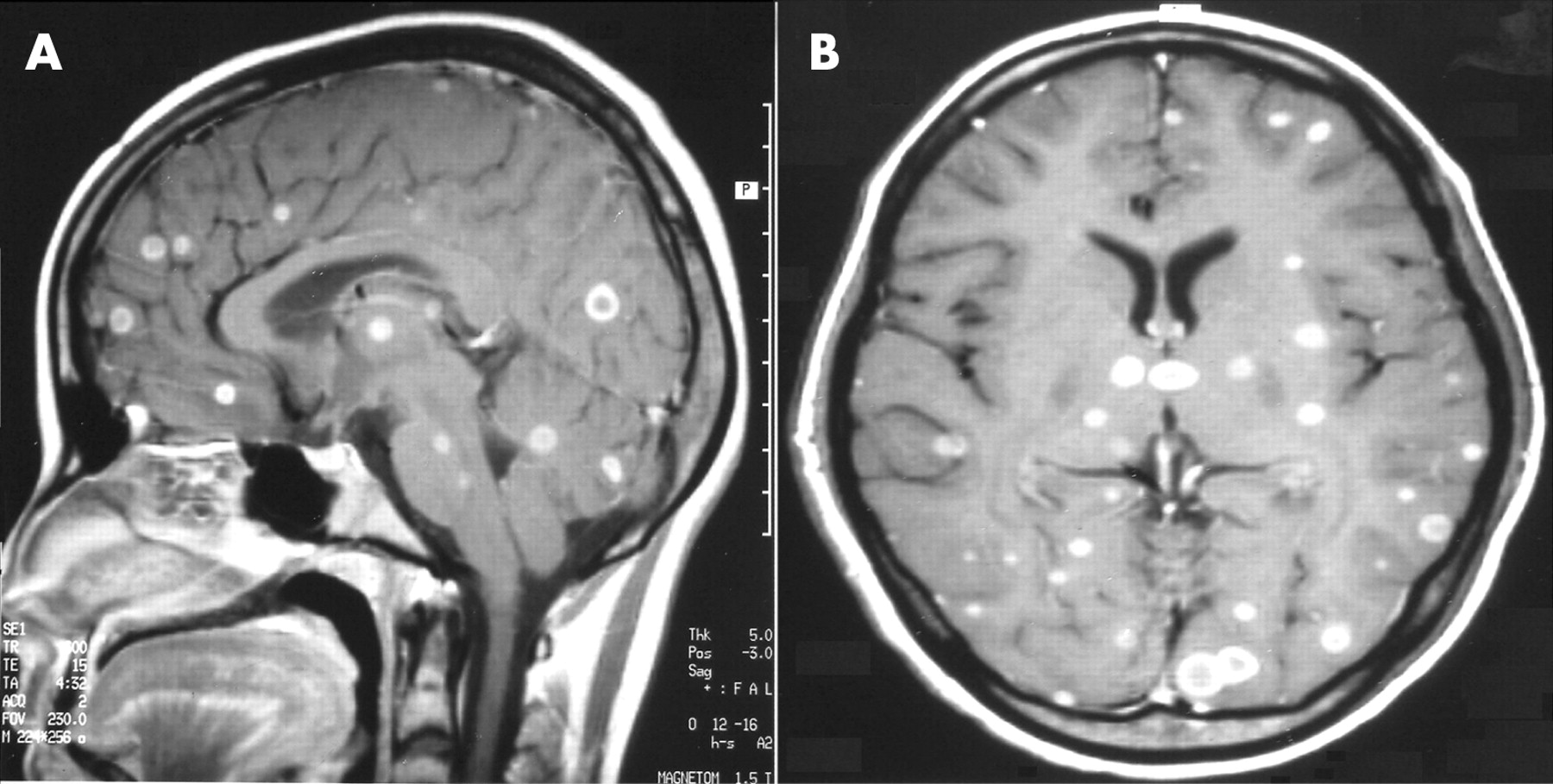
A gadolinium-enhanced MRI scan of the brain showed multiple ring enhancing lesions in bilateral cerebral hemispheres, thalami, basal ganglia, cerebellum and pons (fig 1A, 1B). Some of the lesions were abutting at the surface of brain. No meningeal enhancement was seen. No midline shift was evident.
Further analysis of CSF showed positive PCR for Mycobacterium tuberculosis (Mtb). Tracheal aspirate smear examination by Zeil Nelson’s stain showed Mtb (fig 2). Toxoplasma serology was negative in blood and in CSF. Peripheral lymph node biopsy showed features of reactive lymphadenitis. Ultrasonography of the abdomen was normal. A diagnosis of disseminated tuberculosis with central nervous system (CNS) involvement was made and she started receiving the standard four drug anti-tuberculosis treatment (Isoniazid, Rifampicin, Pyrazinamide and Ethambutol) along with anti-epilepsy drugs (sodium valproate) and supportive care. She started improving and after 4 weeks, highly active antiretroviral therapy consisting of lamivudine, stavudine and efavirenz was started. She recovered, but with residual visual field defects and paraparesis. She has completed 6 months of anti-tuberculosis treatment and is under follow-up with anti-epilepsy drugs and HAART.

Multiple ring-enhancing lesions in HIV positive patients are seen at advanced stages of immunosuppresion. Differential diagnosis primarily includes tuberculoma, primary CNS lymphoma and toxoplasmosis. Sometimes, it may be due to bacterial or fungal abscesses.1–2 In our case, the diagnosis was established by bacteriological proof from two sites. Concurrent chest involvement in disseminated tuberculosis aids in the early diagnosis because of the ease of Mtb detection from sputum, tracheal aspirate or bronchioloalveolar lavage fluid. In HIV positive patients, detection of Mtb may not always be easy because the proportion of extrapulmonary tuberculosis increases and pulmonary disease is of paucibacillary nature.3
Acknowledgments
This article has been adapted from Tahir Mohammad, Das Chandan J, Sharma S K, Sinha Sanjeev, Singh U B. Multiple ring enhancing lesions in brain MRI of a patient with AIDS Journal of Neurology, Neurosurgery and Psychiatry 2007;78:526
Footnotes
Competing interests: None declared.