Article Text

Statistics from Altmetric.com
DESCRIPTION
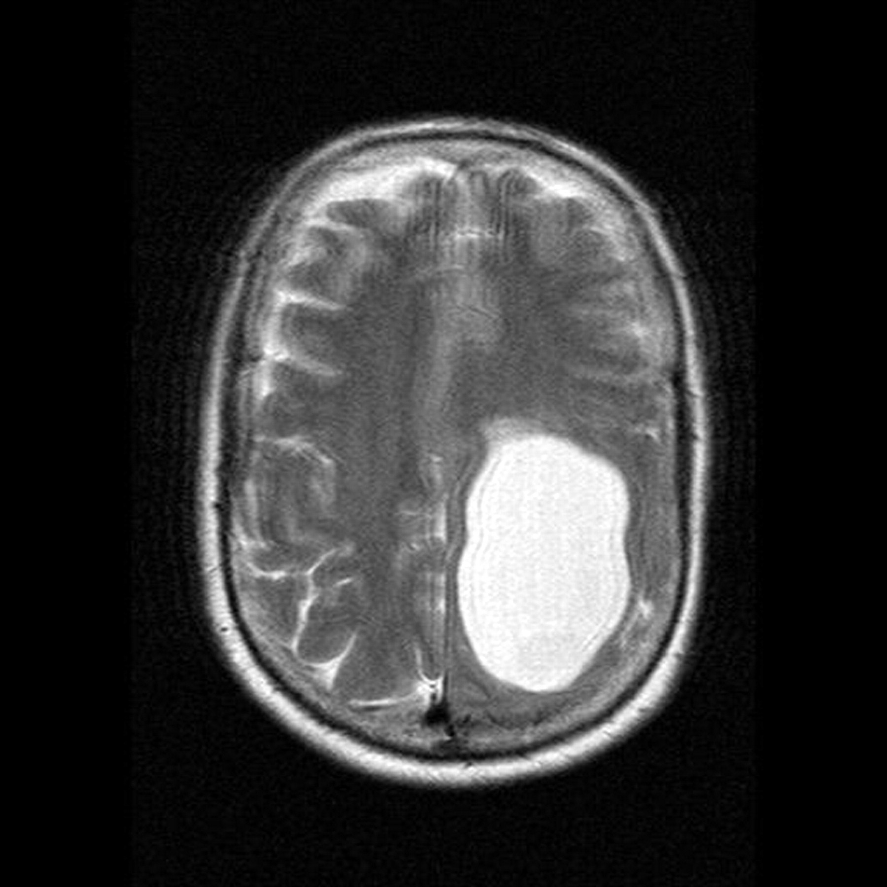
A 57-year-old woman who was right handed presented to a neurologist with day-to-day memory difficulties, forgetting the names of familiar people and objects, and expressive dysphasia. She had been previously fit and well, without any other symptoms. Her physical examination, including the examination of the central nervous system was unremarkable. She scored 23/30 on Mini Mental State Examination losing points for short-term recall, attention and concentration and constructional apraxia. An obvious suspicion was of Alzheimer’s disease;1 however, a magnetic resonance imaging scan was done to rule out a secondary cause of dementia, which showed a huge arachnoid cyst in the left cerebral hemisphere causing mass effect on the left parietal lobe (hence the symptoms). The patient showed significant improvement following surgical treatment. This case elaborates the importance of neuroimaging in all cases of suspected dementia while describing a rare condition.
Arachnoid cysts are formed from a splitting of the arachnoid membrane with an inner and outer leaflet surrounding the cyst cavity.2 They contain cerebrospinal fluid but do not communicate with the ventricular system. The majority are of congenital origin but may develop following meningitis, surgery, trauma, subarachnoid haemorrhage or occur in association with neoplasm. They constitute approximately 1% of intracranial masses with 50–60% occurring in the middle cranial fossa. Cysts in the middle cranial fossa are found more frequently in men and on the left side. Local ischaemia induced by compression due to an arachnoid cyst may cause memory dysfunction and behavioural disturbances.3 Untreated, may cause permanent severe neurological damage due to progressive expansion of the cyst. A variety of neurosurgical procedures may be used to decompress the cyst.
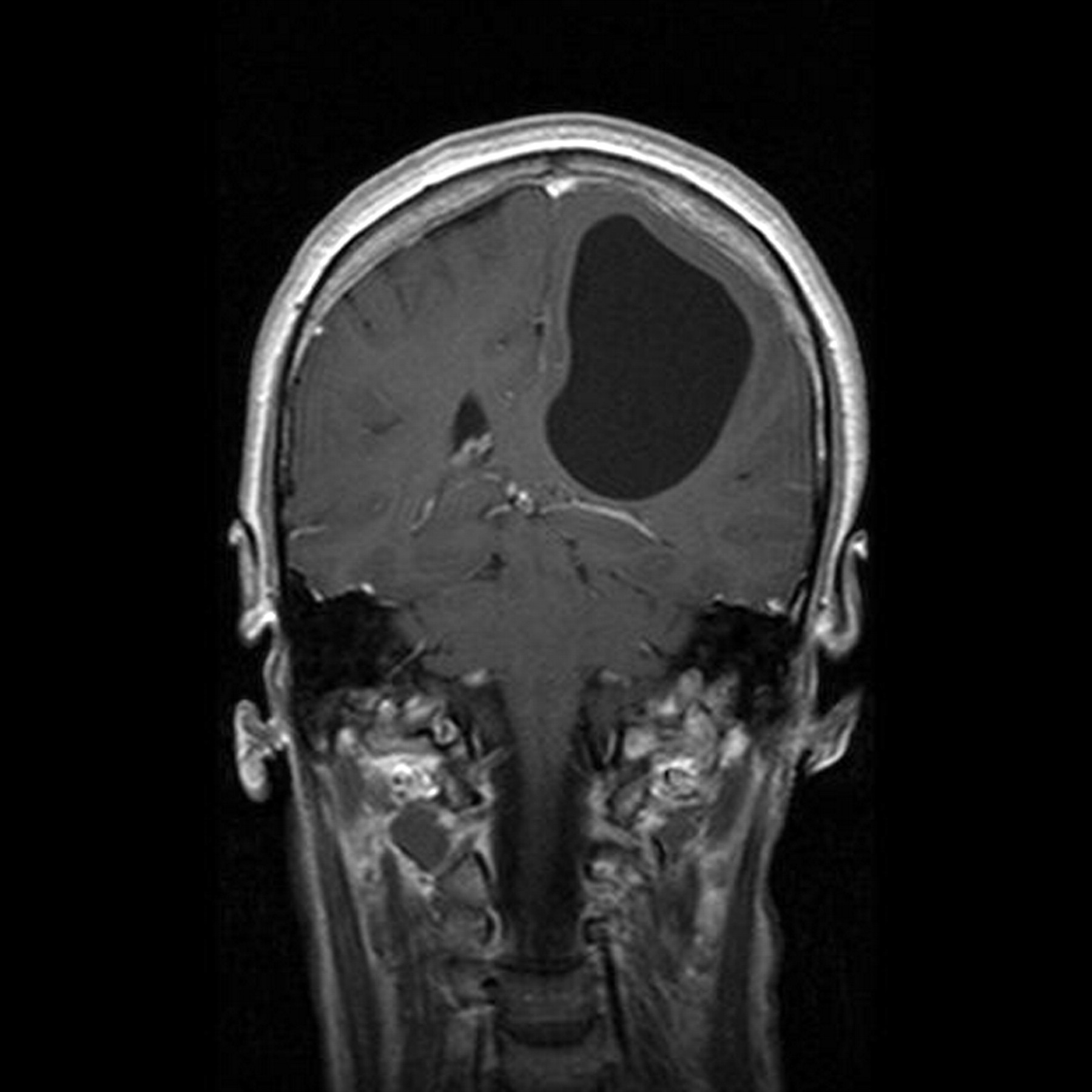
Coronal T1-weighted magnetic resonance imaging scan of the brain showing an arachnoid cyst. The skull vault is normal and there is no evidence of remodelling. Hence the cyst is not long-standing.

Sagittal T2-weighted magnetic resonance imaging scan of the brain showing an arachnoid cyst causing mass effect on the left parietal lobe. The signal in the cyst is exactly the same as cerebrospinal fluid.
Acknowledgments
The patient for giving her consent.
Footnotes
Competing interests: none.
Patient consent: Patient/guardian consent was obtained for publication.