Article Text

Statistics from Altmetric.com
A 28-month-old girl presented with lethargy and unable to bear weight. She was severely anaemic and had a large right sided abdominal mass in lumbar area that was bimanually palpable and ballotable. She was normotensive and systemic examination was otherwise normal. Preliminary investigations showed severe microcytic hypochromic anaemia with haemoglobin of 3.9 gm/dl, neutrophilic leucocytosis and normal platelets. Renal profile, liver enzymes, creatine kinase, thyroid function and uric acid values were normal. C reactive protein (CRP) (238 mg/l), erythrocyte sedimentation rate (ESR) (162 mm/h) and immunoglobulins were high. Urine microscopy showed 10 white cells, no red cells. Urine and blood cultures were negative. Stool occult blood was negative.
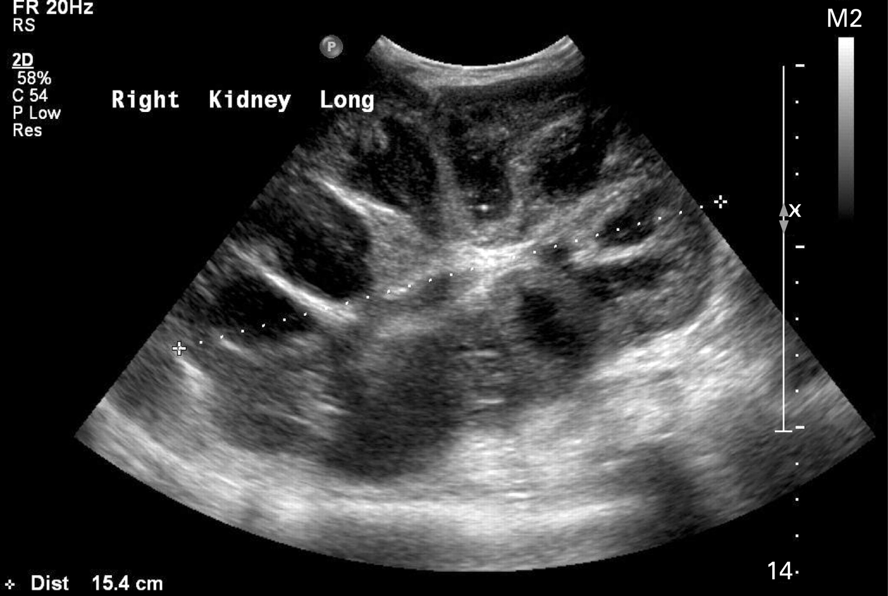
Renal ultrasound scan suggested right renal or suprarenal mass without acute urinary obstruction (fig 1).

Ultrasonographic appearance of abnormal right kidney. Right kidney measured 15.4 cm with distinctive low echogenicity areas centrally without definite communication with renal pelvis. Two areas of focal echogenicity consistent with renal calculi were identified; one at the renal pelvis and the other at the lower pole.
Bone Marrow was normocellular but showed reduced erythropoiesis.
Computed tomography (CT) scan with contrast confirmed the ultrasonographic findings and also showed a non-functional right kidney (fig 2). The appearance of the right kidney with significant calyceal dilatation, foci of calcification at the periphery with a large calculus at renal pelvis suggested xanthogranulomatous pyelonephritis (XGP).

Computed tomography scan of abdomen with contrast, showing non-functional, abnormally enlarged right kidney with calculus, while the left kidney takes up contrast.
Right nephrectomy was performed by transperitoneal approach and the diagnosis was confirmed (fig 3). Microscopically sheets of foamy histiocytes along with plasma cells, lymphocytes and neutrophilic exudates were seen without any granulomata or neoplasia. Pus from the specimen grew Proteus species.

Right nephrectomy specimen: normal renal architecture being replaced by multiple cystic spaces near the hilum filled with necrotic pus-like material with solid nodular appearances at the periphery.
Malignant tumours and chronic blood loss are well known to cause severe anaemia in children. This child had XGP, a rare form of locally invasive chronic atypical inflammatory renal disease resulting in severe iron deficiency anaemia without any evidence of blood loss or neoplastic bone marrow involvement. XGP accounts for 6/1000 surgically proven cases of chronic pyelonephritis.1 The exact aetiology is unknown, but it is generally accepted that the disease process requires long term renal obstruction and infection. Various aetiologies have been suggested such as calculus or non-calculus urinary obstruction, ineffectively treated urosepsis, local alteration in renal metabolism or perfusion, lymphatic obstruction, lipid metabolism alterations, or altered immune responses.
Focal XGP may be hard to differentiate from renal parenchymal tumours.1 The characteristics that might help in preoperative diagnosis based on clinical observations2 include unilateral renal disease with grossly impaired function, large (often numerous) renal calculi in association with anaemia, raised ESR, and leucocytosis. Radiological investigations are extremely useful in the diagnosis, but no single clinical or radiological sign is pathognomonic3 and diagnosis has to be confirmed histologically.
XGP is rare in children and its presentation can be atypical. Clinical diagnosis can be challenging. In the case reported here, the child remained apyrexial and had no clinical or laboratory evidence of urinary tract infection, though she had an unobstructed urinary tract with severe chronic pyelonephritis. She therefore presented late to hospital with anaemia and lethargy. Her severe anaemia could be due to a combination of chronic infection and nutritional deficiency. The manifestations of XGP mimic neoplastic and other inflammatory renal parenchymal diseases, including tuberculosis, and is often misdiagnosed preoperatively. Increased awareness about this condition could improve the chances for proper preoperative diagnosis and management, resulting in a better long term outcome.
Acknowledgments
We acknowledge the contributions of Dr T Anbu, consultant paediatrician who was actively involved in the clinical care, Dr Caren Landes, consultant radiologist, for radiological diagnosis and interpretation of images, and Dr Rajeev Shukla, consultant pathologist for histopathological diagnosis and guidance.
Footnotes
Competing interests: none.
Patient consent: Patient/guardian consent was obtained for publication