Article Text

Statistics from Altmetric.com
DESCRIPTION
A 16-month-old boy presented with developmental regression. He had previously been completely well and had achieved his developmental milestones appropriately until the age of 14 months. At this point his development started to regress and his parents noted a change in his personality. He had been born at term by normal vaginal delivery with no neonatal problems. The pregnancy was unremarkable, as were the antenatal scans.
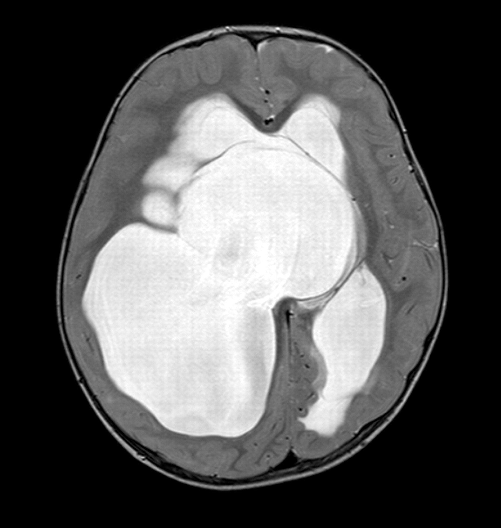
When we reviewed him, his occipitofrontal circumference (OFC) was 54.3 cm (>99.6th percentile). He was irritable, floppy and unable to sit without support. An urgent MRI scan (fig 1) showed a large multiloculated cyst, which originated in the right cerebral hemisphere and extended across the midline and into the posterior fossa. The cyst compressed both lateral ventricles and the white matter, the ventricles were dilated. There was evidence of periventricular high signal changes indicating oedema secondary to the hydrocephalus. A diagnosis of complex multiloculated cystic hydrocephalus was made.
MRI scan (axial T2-weighted) of brain showing complex multiloculated cystic hydrocephalus.
The child was transferred to the regional paediatric neurosurgical department and underwent endoscopic fenestration of the cyst. He was discharged 2 days post-operatively. Follow-up showed that his OFC had stabilised and he had started to regain developmental milestones, including walking. A repeat MRI scan showed significant reduction in the size of the cysts and the lateral ventricles.
Multiloculated hydrocephalus is a rare form of hydrocephalus in which the presence of septations or obstructions inside the ventricular system leads to accumulation of fluid in isolated compartments. It is usually related to previous intracranial haemorrhage or infection, but can be idiopathic. Urgent investigation and neurosurgical intervention is required.1,2
Footnotes
Competing interests: none.
Patient consent: Patient/guardian consent was obtained for publication.