Article Text

Summary
A 27-year-old competitive surfer presented with a history of a painful right ankle. He was able to recall an injury to his right ankle 4–5 years previously, sustained while surfing. The mechanism described was that he had dropped a considerable height during take-off, sustaining an impact injury from the board. He recalled immediate pain and swelling followed by 2–3 weeks of pain and a limp; he continued to surf, albeit with difficulty, despite this.
Investigations found him to have a bony spur on the anterolateral part of the talus. This case shows how this injury is similar to those observed in other sports.
Statistics from Altmetric.com
BACKGROUND
Surfing is a popular sport in coastal regions of the UK. Situated close to a popular surfing area, our department treats a number of acute and chronic surfing injuries. We describe a chronic ankle injury detected in a high level surfer that posed a diagnostic problem. An increasing awareness of these conditions will direct appropriate investigation and management and expedite the return of these patients to their desired/required level of activity.
CASE PRESENTATION
A 27-year-old competitive surfer presented with a history of a painful right ankle. He was able to recall an injury to his right ankle 4–5 years previously sustained while surfing. The mechanism described was that he had dropped a considerable height during take-off, sustaining an impact injury from the board. He recalled immediate pain and swelling followed by 2–3 weeks of pain and a limp; he continued to surf, albeit with difficulty, despite this.
He developed gradually increasing symptoms of pain and an audible and palpable click in the anterolateral aspect of his right ankle. He noticed that he had lost movement in his ankle such that it was limiting daily activities and preventing him from competitive surfing.
On clinical examination his gait appeared normal. There was a palpable swelling and tenderness in the region of the anterior talofibular ligament. Dorsiflexion at the ankle joint was reduced to only 5°, with full plantar flexion and no evidence of instability.
INVESTIGATIONS
Plain radiographs, weight bearing views and stress views were normal. An MRI scan was reported as normal. A subsequent ultrasound scan demonstrated a bony spur from the superolateral aspect of the talus with “multiple osseous bodies”. A CT scan was performed to further delineate the bony abnormality and confirmed the osseous spur (fig 1A,B) and associated calcification of the anterolateral talus with impingement of the extensor digitorum tendons.
Dimensional CT reconstruction of the ankle with an obvious talar bony spur.
TREATMENT
A right ankle arthrotomy and excision of the bony lesion was performed. Histopathology confirmed the pathology was in keeping with the impression of a posttraumatic lesion
OUTCOME AND FOLLOW-UP
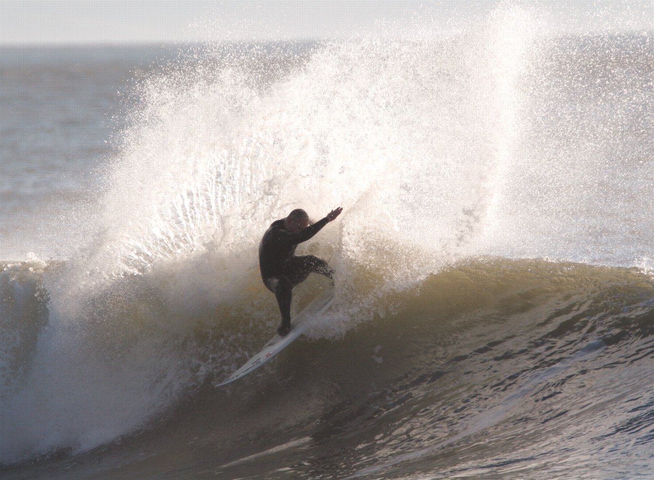
The patient underwent a period of extensive rehabilitation with physiotherapy. The physiotherapy exercises consisted of specific exercises designed to increase the range of motion of the ankle, concentric and eccentric exercises designed to improve the power of the ankle muscles, gait re-education progressing from walking aids to running, static and dynamic proprioceptive ankle exercises and plymoteric exercises progressing from theraband to single-leg hoops. He returned to competitive surfing, becoming the Welsh Open surfing champion the following year and at 2 years later has no further symptoms (fig 2).

The patient back in competitive surfing.
DISCUSSION
Articles devoted to surfing injuries in the medical literature are scarce. A study by Nathanson et al1 discovered that fractures accounted for 8% of surfing injuries and 37% of all surfing injuries were to the lower limbs. They found that the majority of injuries were caused by the surfer’s own board. They did not comment on any significant ankle injuries.
Zoltan et al2 determined that surfers were most likely to be injured by their own board. They commented that protective shielding either on the board or surfer might prevent some of the injuries. They found that the use of the leash reduced the injury caused by stray boards hitting surfers.
A review of professional surfing injuries sustained in competitions from 1999 to 2005 found that the size of the wave and type of sea bed being surfed over increased the risk of injury to the surfer.3
Imaging of the injured ankle can sometimes be problematic, as illustrated by this case. MRI is widely accepted as the modality of choice when it comes to acute ankle injuries. This is because it allows bone and soft tissue to be imaged in detail.4 The utilisation of defined MRI sequences has resulted in an increased sensitivity and specificity for accurate diagnoses when compared to normal MRI scans of the ankle.5 Plain radiographs may offer some value in the diagnosis of bony spurs in cases of ankle impingement. In patients who have anteromedial impingement, these radiographs can often be falsely negative. Oblique plain films used in a study by, Van Dijk et al6 found that they added little in the diagnosis of anterolateral spurs compared to normal lateral views. There is evidence that ultrasound can be beneficial in tiny fractures of the ankle as a result of “ankle sprain”.7,8 It is important to note that these studies looked primarily at acute ankle injuries while the injury in this case report is a chronic injury.
A study from The Netherlands9 suggests that the distal fascicle of the anterior inferior tibiofibular ligament as a cause of tibiotalar impingement syndrome should be considered as a cause for impingement in the stable ankle joint with normal radiographical findings.
The so-called “snowboarder’s ankle” is thought to be due to an injury to the lateral talus, which may be similar to this case although the mechanism of injury is different to that sustained by the surfer in our case. One hypothesis for this is the fact that snowboarding boots are softer than those worn while skiing. A study by Delorme et al10 confirmed that using stiffer boots reduced the amount of movement at the ankle joint.
This case illustrates the importance of equating mechanism of injury to pathology in terms of aetiology and the forces and demands upon a surfer’s ankle. It also demonstrates the importance of combining multiple imaging modalities in terms of achieving a diagnosis; relying heavily on a single modality may not be sensitive or specific enough in cases such as these.
LEARNING POINTS
-
It is important to think of the mechanism of injury.
-
There is a need to consider the benefits of various modalities of investigation.
-
Clinical examination is important in any investigation.
REFERENCES
Footnotes
Competing interests: None.
Patient consent: Patient/guardian consent was obtained for publication.