Article Text

Statistics from Altmetric.com
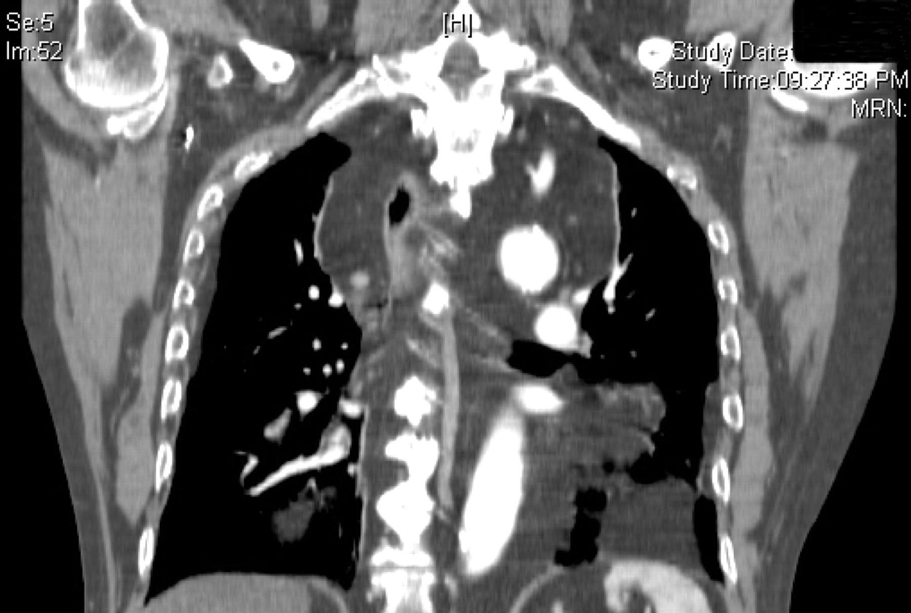
A 64-year-old man with known inflammatory bowel disease, on corticosteroid treatment, presented with drowsiness and poorly defined central chest discomfort. As part of his routine work up he underwent a chest x ray (CXR) and 12 lead electrocardiogram (ECG). Though the ECG was normal, the CXR demonstrated a grossly widened mediastinum (fig 1). The possibility of an acute aortic dissection was raised and an urgent computed tomography (CT) aortogram was performed. Though the CT scan was negative for an aortic lesion, it demonstrated the presence of pronounced mediastinal lipomatosis (fig 2).


Mediastinal lipomatosis is a benign condition characterised by symmetric accumulation of fat within the mediastinum, which distorts the mediastinal silhouette by varying degrees. It is a distinct clinical entity that occurs with systemic corticosteroid treatment, simple obesity or Cushing’s syndrome.
One should consider the diagnosis of mediastinal lipomatosis in patients whose clinical history is not consistent with alternative diagnoses of mediastinal widening (aortic dissection, mediastinal haemorrhage, mediastinitis). In such patients mediastinal lipomatosis should be suspected if: (1) the mediastinal widening is bilateral; (2) the contour is smooth and sharply defined; (3) the radiolucency is not as pronounced as surrounding structures; and (4) where there is an absence of tracheal compression.1
REFERENCES
Footnotes
Competing interests: None.
Patient consent: Patient/guardian consent was obtained for publication