Article Text

Summary
Retinal artery occlusions are usually the result of emboli, although non-embolic causes such as vasculitides, coagulopathies, and vasospasms resulting from migraines and inflammatory conditions do occur. Bevacizumab, a humanised monoclonal antibody, is designed to bind to and inhibit vascular endothelial growth factor (VEGF). VEGF is a protein that plays a critical role in tumour angiogenesis, the formation of new blood vessels to the tumour. The use of bevacizumab has spread worldwide, but the drug related adverse events associated with its use have been reported in a few retrospective reviews. We report on a previously unpublished case of retinal artery occlusion following the use of systemic bevacizumab.
Statistics from Altmetric.com
BACKGROUND
We report on a previously unpublished case of retinal artery occlusion following the use of systemic bevacizumab. Before this report, several studies have shown evidence of systemic thromboembolic events after the use of intravenous bevacizumab, but until now no case has been reported of retinal artery occlusion immediately after administration of systemic bevacizumab.
CASE PRESENTATION
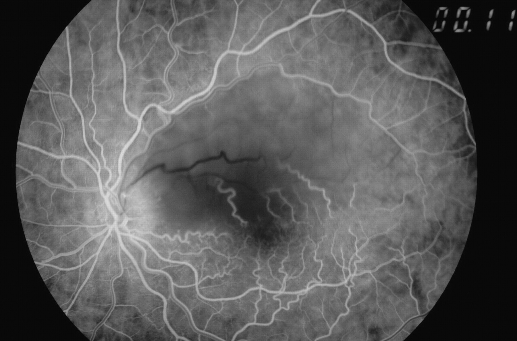
A 45-year-old male patient with lung cancer noticed procursive blurring of the inferior visual field in the left eye 3 days after intravenous bevazicumab administration as a part of his chemotherapy. He presented to our clinic 1 week after the ocular symptoms had started. He was diagnosed with stage III non-small cell carcinoma of the lung in 2008 and had since been treated with intravenous bevacizumab 15 mg/kg plus up to six cycles of chemotherapy with carboplatin and paclitaxel (200 mg/m) every 3 weeks. Examination showed the patient to be afebrile with a blood pressure of 161/85 mm Hg. The patient had a normal complete blood cell count and all coagulation factors assayed in the patients were within the normal ranges. The patient was referred for a detailed cardiac and carotid evaluation including carotid Doppler study and echocardiography which was unremarkable. There was no evidence of metastasis to bone or brain. Best corrected visual acuity was 20/100 in left eye, 20/20 in right eye. On slit lamp examination, the anterior segment was unremarkable in both eyes; no anterior vitreous cells or flare was observed. Intraocular pressure was 16 mm Hg bilaterally. His left eye showed typical findings of branch retinal artery occlusion (BRAO). Fundus examination of the left eye showed a patchy area of partially faded retinal whitening in the superotemporal quadrant of the macula, just involving the fovea, with attenuation and sclerosis of the macular division of the superotemporal branch retinal artery (fig 1). The right fundus was normal. Early phase fluorescein angiogram of the left eye showed narrowing of the affected arterial segment, blocked background fluorescence in the area of whitening, and delayed arteriovenous transit in the affected vessels (fig 2). Goldmann kinetic visual field analysis disclosed an inferonasal field defect corresponding to the area of arterial occlusion. He was followed up for 3 months. His visual acuity had improved to 20/50, but the inferior field defect persisted in the left eye.
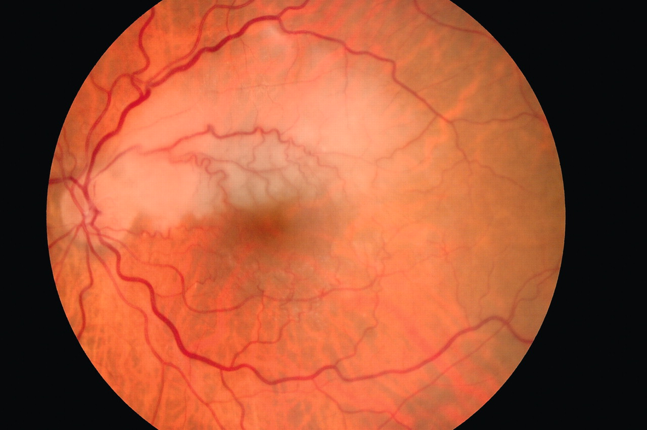
Fundus view of left eye showing superficial retinal whitening with the occluded artery in the superotemporal quadrant of the macula.
Early phase fluorescein angiogram of the left eye shows narrowing of the superiotemporal branch retinal artery, blocked background fluorescence in the area of whitening, and delayed arteriovenous transit in the affected vessels.
TREATMENT
Based on the duration of the patient’s visual loss, the opinion was that any therapeutic intervention would be unlikely to improve vision. Nevertheless, prophylactic warfarin was started by his oncologist to prevent thromboembolic events, with a target international normalised ratio (INR) of approximately 2.
OUTCOME AND FOLLOW-UP
The patient was followed up for 3 months. His visual acuity had improved to 20/50, but the inferior field defect persisted in the left eye.
DISCUSSION
Arterial thrombosis and occlusion is considered a disease of the elderly, with more than 90% of cases occuring in patients >50 years of age, with associated systemic vascular disease and vascular endothelial damage.1–3 Risk factors predisposing patients to BRAO include hypertension, endothelial cell damage, carotid occlusive disease or atherosclerosis, coronary artery disease, and hypercholesterolaemia. Bevacizumab was approved by the US Food and Drug Administration in February 2004 for use as part of combination therapy for metastatic colorectal cancer. The results from clinical studies of bevacizumab as a single agent or as part of combination regimens for breast cancer, non-small cell lung cancer, renal cell carcinoma, and other solid malignancies have been promising.4,5 Although combination treatment with bevacizumab and chemotherapy improves survival of patients with various carcinomas, an increased risk of arterial thromboembolic events has been observed.6 The vast majority of the vascular occlusions have been attributed to degenerative changes of the vessel wall.2,3 Antagonising vascular endothelial growth factor (VEGF) as a major endothelial mitogen might decrease the renewal capacity of the endothelial cell in response to trauma and result in endothelial dysfunction. VEGF antagonism could also cause decreased matrix deposition in the supporting layers of vessels. Therefore, anti-VEGF therapy might cause not only a tendency to bleed due to a decreased renewal capacity of endothelial cells, but an increased risk of thrombotic events that could also be encountered as a result of tissue factor activation secondary to exposure to subendothelial collagen and endothelial damage.6
Michel et al showed that intravenous administration of bevacizumab produced significant improvement in visual acuity and reduction of central retinal thickening in patients with age related macular degeneration (ARMD) without any ocular side effect.7 Off-label intravitreal injections of bevacizumab have been given for the treatment of neovascular and exudative ocular diseases since May 2005. However, the treatment of intravitreal injection is also not without risks. The incidence of central retinal artery occlusion possibly related to intravitreal bevacizumab was reported at 0.01%.5 Intravitreal bevacizumab has been used in treatment of the complications of retinal artery occlusion.8 Before this report, several studies have shown evidence of systemic thromboembolic events after the use of intravenous bevacizumab, but no case has reported retinal artery occlusion immediately after administration of systemic bevacizumab. Ophthalmologists and oncologists should therefore be aware of this rare critical ocular complication following intravenous or intravitreal bevacizumab injection.
LEARNING POINTS
-
A case of retinal artery occlusion following the use of systemic bevacizumab is reported.
-
Ophthalmologists and oncologists should be made aware of this rare critical ocular complication following intravenous or intravitreal bevacizumab injection.
Footnotes
Competing interests: none.
Patient consent: Patient/guardian consent was obtained for publication