Article Text

Statistics from Altmetric.com
Description
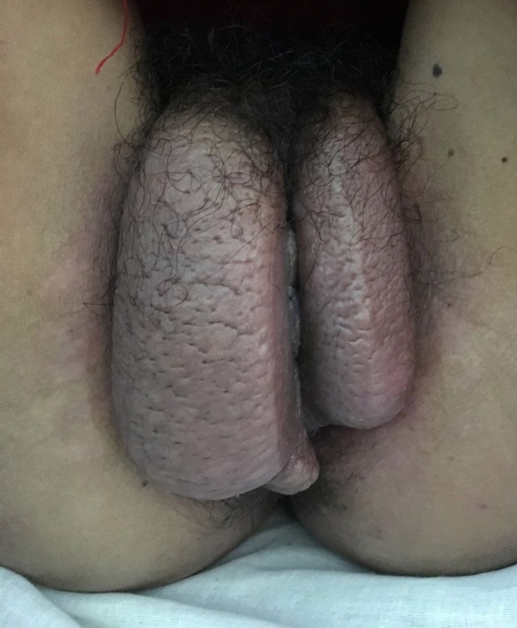
A 25-year-old unmarried woman, resident of a filarial endemic region, presented with progressive swelling of the vulva along with recent onset redness, dragging pain and consequent difficulty in walking. She had noticed the vulval swelling about 2 years before her presentation and had been suffering from recurrent episodes of low-grade fever for the preceding 2 years. The swelling had been reducible initially and became non-reducible after about 6 months. She denied any pelvic surgery, radiation or trauma. Her medical and family histories were unremarkable. A thorough systemic examination did not document any abnormality. Genital examination revealed non-ulcerative bilateral vulval swelling with bosselated surface, hard in consistency with raised local temperature and palpable bilateral superficial inguinal lymph nodes (figure 1). Baseline work-up was as follows: haemoglobin 11 g/dL, total leucocytes count 7000 cells/mm3, neutrophil 60%, lymphocytes 20%, eosinophil 12% and erythrocyte sedimentation rate (ESR) 60 mm in first hour. Peripheral blood collected at midnight examined by concentration technique using Leishman stain was negative for microfilaria. However, immunochromatographic test for microfilarial antigen was positive. Mantoux test was negative, chest X-ray was normal and pelvic ultrasonography was unremarkable. Both fine needle aspiration cytology and punch biopsy from the involved area showed features of chronic inflammation as evidenced by perivascular collection of lymphocytes, plasma cells, and eosinophils and occasional foreign body granuloma without acid fast bacilli in Ziehl–Neelsen stain, fungal hyphae in Periodic acid–Schiff stain or Donovan bodies or Chlamydia elementary bodies in Giemsa stain. The woman was treated with diethylcarbamazine (DEC) in three divided doses of 6 mg/kg/day for 21 days and was advised to wear tight undergarments and to maintain local hygiene to prevent any secondary infection. Cosmetic surgery was advised after completion of the course of DEC, and the woman was referred to the department of plastic surgery.
Bilateral vulval swelling.
Lymphedema is swelling of soft tissue that develops following accumulation of protein-rich interstitial fluid secondary to improper lymphatic drainage of the involved area. If untreated for prolonged period, lymphatic obstruction can ultimately result in massive lymphedema, also known as elephantiasis, and vulval elephantiasis accounts for only about 1%–2% of total elephantiasis cases. Infections like filariasis, tuberculosis, lymphogranuloma venereum, donovanosis and a number of non-infective conditions such as genital malignancies, metastatic Crohn’s disease, inguinal and pelvic lymph node dissection, genital radiation, vulvectomy or pelvic trauma can at times give rise to genital elephantiasis.1 Genital elephantiasis, also known as esthiomene, is rare; even rarer is genital elephantiasis secondary to filarial infections, seen in only 1%–2% of all such cases.2 Lymphatic filariasis is caused by Wuchereria bancrofti and Brugia malai. The third-stage microfilarias after entering the human body by bite of a mosquito reside within the lymphatic system and begin to grow into adult forms. After acquiring sexual maturity, the male fertilises the female to give birth to the larva. The lymphatic damage is caused by the movement of the adult worm producing inflammatory reactions and resultant lymphangitis, leading to stasis of lymph and subsequent lymphedema. To overcome the stasis, affected lymphatics undergo dilatation, become tortuous and a number of collateral lymph vessels develop. Further movement of adult worm ultimately leads to hypertrophy and hyperplasia of lymphatic endothelium which further aggravates stasis. Thus with time the lymphatic obstruction becomes irreversible and leads to elephantiasis. It is postulated that as long as the adult worms are alive, the lymphatic vessels remain patent. Death of the worm, however, leads to extensive perilymphangitis, progressive fibrosis and obliteration of vessels.3 Therefore, the worm is usually not found in peripheral blood or within the involved tissue in these patients and clinical suspicion along with ancillary tests are the cornerstones of diagnosis.
In addition to DEC that kills both the adult worms and microfilaria, maintenance of local hygiene, proper skin care and prompt treatment of secondary bacterial infections, if any, are of utmost importance. However, majority require cosmetic surgical interventions like excision therapy with or without split thickness skin graft or thigh flaps. Though uncommon, recurrence after initial reconstructive surgery may also be encountered.
Learning points
Though vulval elephantiasis due to filarial infection is very rare, in endemic countries it should be considered as one of the important causes of elephantiasis, both limb and genital.
Vulval filariasis is essentially a clinical diagnosis. In the absence of documentation of filarial worm in peripheral smear or within the involved tissue section, elevated absolute eosinophil count, positive filarial antigen assay and ruling out other possible differentials establish the diagnosis.
Genital elephantiasis significantly affects the physical, mental and social well-being of a patient; so early diagnosis and effective treatment of filariasis is the only key to avoid such physical disability and extreme mental anguish.
Aims of the management of vulval filariasis are reduction of vulval swelling, prevention of secondary infections and restoration of the normal genital appearance and sexual functions. Diethylcarbamazine with or without antibiotics and cosmetic surgery are the available therapeutic options.
Footnotes
Contributors All authors were involved in diagnosis and management of the patient. MC and HB did the literature search. PPC wrote the manuscript.
Competing interests None declared.
Patient consent Obtained.
Provenance and peer review Not commissioned; externally peer reviewed.